Click the link to purchase on Amazon 🎉📚
==============================================
🎥 Check Out All Videos at Once! 📺
👉 Visit Visualizing MSK Blog to explore a wide range of videos! 🩻
https://visualizingmsk.blogspot.com/?view=magazine
📚 You can also find them on MSK MRI Blog and Naver Blog! 📖
https://www.instagram.com/msk_mri/
Click now to stay updated with the latest content! 🔍✨
==============================================
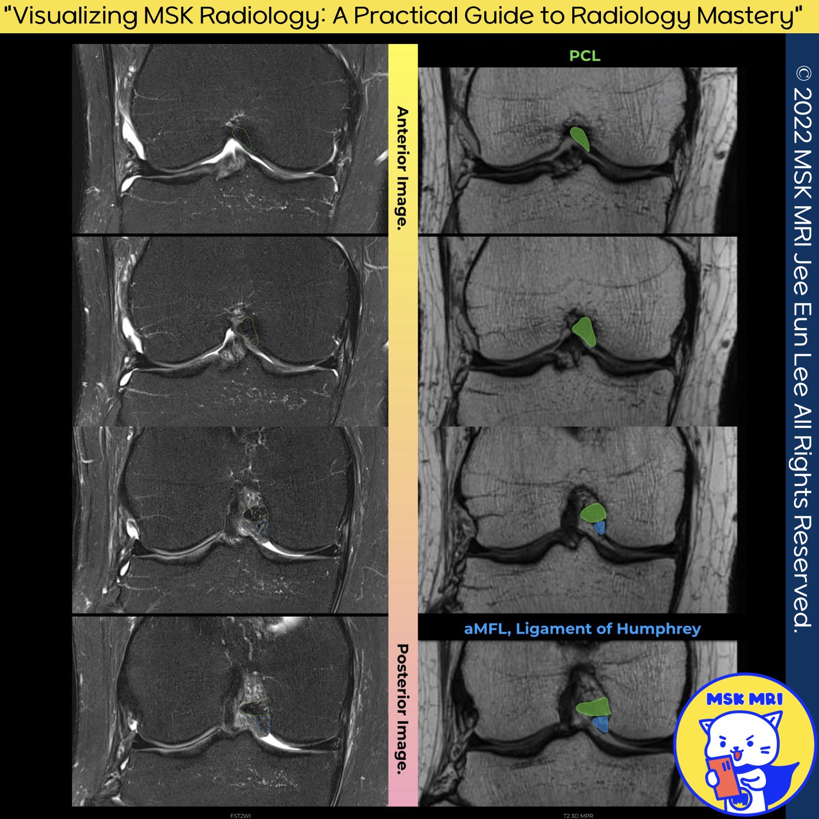
1️⃣ Anatomy of the Proximal PCL on Coronal Images:
- The proximal PCL appears more circular in cross-section on mid- and anterior-coronal images.
- On the posterior coronal plane, the PCL shows a more vertical orientation. This is because it's sectioned along the downward slope of its curved arc.
Stoller's Orthopaedics and Sports Medicine: The Knee
2️⃣ Meniscofemoral Ligaments (MFLs):
- The presence of the anterior and posterior MFL varies among individuals. The anterior MFL is observed in 20–75% of knees, whereas the posterior MFL appears in 70–100% of cases. Overall, at least one type of MFL is present in more than 90% of individuals.
- The MFLs attach near the Posterior Medial Bundle's (PMB) femoral attachment. Specifically, the posterior MFL attaches just proximal to it, and the anterior MFL attaches just distal, both exhibiting a circular cross-section.
Am J Sports Med. 2015 Dec;43(12):3077-92
3️⃣ Details on Meniscofemoral Ligaments:
- The attachment site of the anterior MFL (aMFL) varies, with 80% attaching distally to the PMB and the remaining 20% to the Anterolateral Bundle (ALB).
- The posterior MFL (pMFL) has a significant femoral attachment area, located proximal to the medial intercondylar ridge and PMB, and attaches distally to the posterior horn of the lateral meniscus.
Am J Sports Med. 2015 Dec;43(12):3077-92
https://visualizingmsk.blogspot.com/?view=flipcard
Visualizing MSK Radiology
visualizingmsk.blogspot.com
"Visualizing MSK Radiology: A Practical Guide to Radiology Mastery"
© 2022 MSK MRI Jee Eun Lee All Rights Reserved.
#VisualizingMSK #PCLinjuries #KneeMRI #PCLtear #PCLanatomy #kneeanatomy
You should not distribute or commercially exploit the content.
You should not transmit or store it on any other website or electronic retrieval system.