Click the link to purchase on Amazon 🎉📚
==============================================
🎥 Check Out All Videos at Once! 📺
👉 Visit Visualizing MSK Blog to explore a wide range of videos! 🩻
https://visualizingmsk.blogspot.com/?view=magazine
📚 You can also find them on MSK MRI Blog and Naver Blog! 📖
https://www.instagram.com/msk_mri/
Click now to stay updated with the latest content! 🔍✨
==============================================
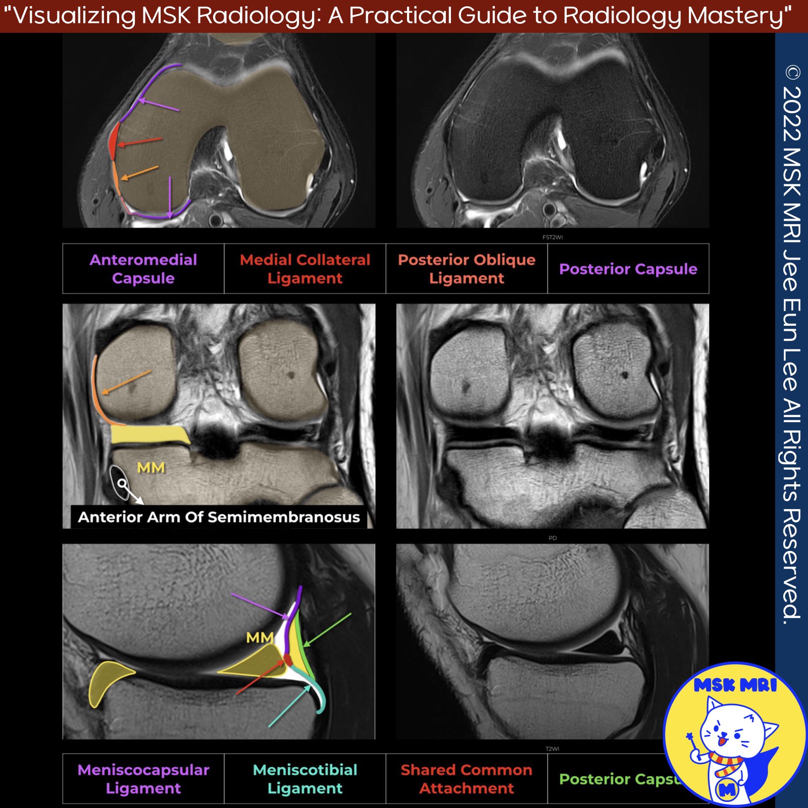
📌 MRI Anatomy of the Posterior Oblique Ligament
✅ Proximal Attachments
The posterior oblique ligament (POL) originates near the adductor tubercle from the posterior margin of the superficial medial collateral ligament (sMCL), where the sMCL fuses with the deep medial collateral ligament.
Visualization The POL is best visualized on coronal and axial MRI sequences. Proximally, it can be identified immediately posterior to the sMCL. However, more distally it becomes difficult to separate from the posteromedial capsule.
✅ Distal Attachments
The main distal attachments of the POL include:
- Tibia
- Posterior horn of the medial meniscus
- Sheath of the semimembranosus tendon
- Posteromedial joint capsule
✅ Three main arms or components:
- Capsular arm - Blends with posteromedial capsule
- Central arm - Inserts into medial meniscus and adjacent tibia
- Superficial arm - Extends superficial to semimembranosus tendon
"Visualizing MSK Radiology: A Practical Guide to Radiology Mastery"
© 2022 MSK MRI Jee Eun Lee All Rights Reserved.
No unauthorized reproduction, redistribution, or use for AI training.
#PMC, #POL, #Posteriorobliqueligament, #kneeMRI, #Kneeanatomy, #anatomyKnee,


'✅ Knee MRI Mastery > Chap 3.Collateral Ligaments' 카테고리의 다른 글
| (Fig 3-A.29) Grade I Posterior Oblique Ligament Injury (0) | 2024.05.11 |
|---|---|
| (Fig 3-A.28) Anteromedial Rotatory Instability (0) | 2024.05.10 |
| (Fig 3-A.26) Three arms of the posterior oblique ligament, anatomy (0) | 2024.05.10 |
| (Fig 3-A.25) Reverse Segond Fracture (0) | 2024.05.09 |
| (Fig 3-A.24) Meniscotibial Ligament Tear_ Part 2 (0) | 2024.05.09 |