Click the link to purchase on Amazon 🎉📚
==============================================
🎥 Check Out All Videos at Once! 📺
👉 Visit Visualizing MSK Blog to explore a wide range of videos! 🩻
https://visualizingmsk.blogspot.com/?view=magazine
📚 You can also find them on MSK MRI Blog and Naver Blog! 📖
https://www.instagram.com/msk_mri/
Click now to stay updated with the latest content! 🔍✨
==============================================
출처: https://mskmri.tistory.com/1056 [MSK MRI:티스토리]
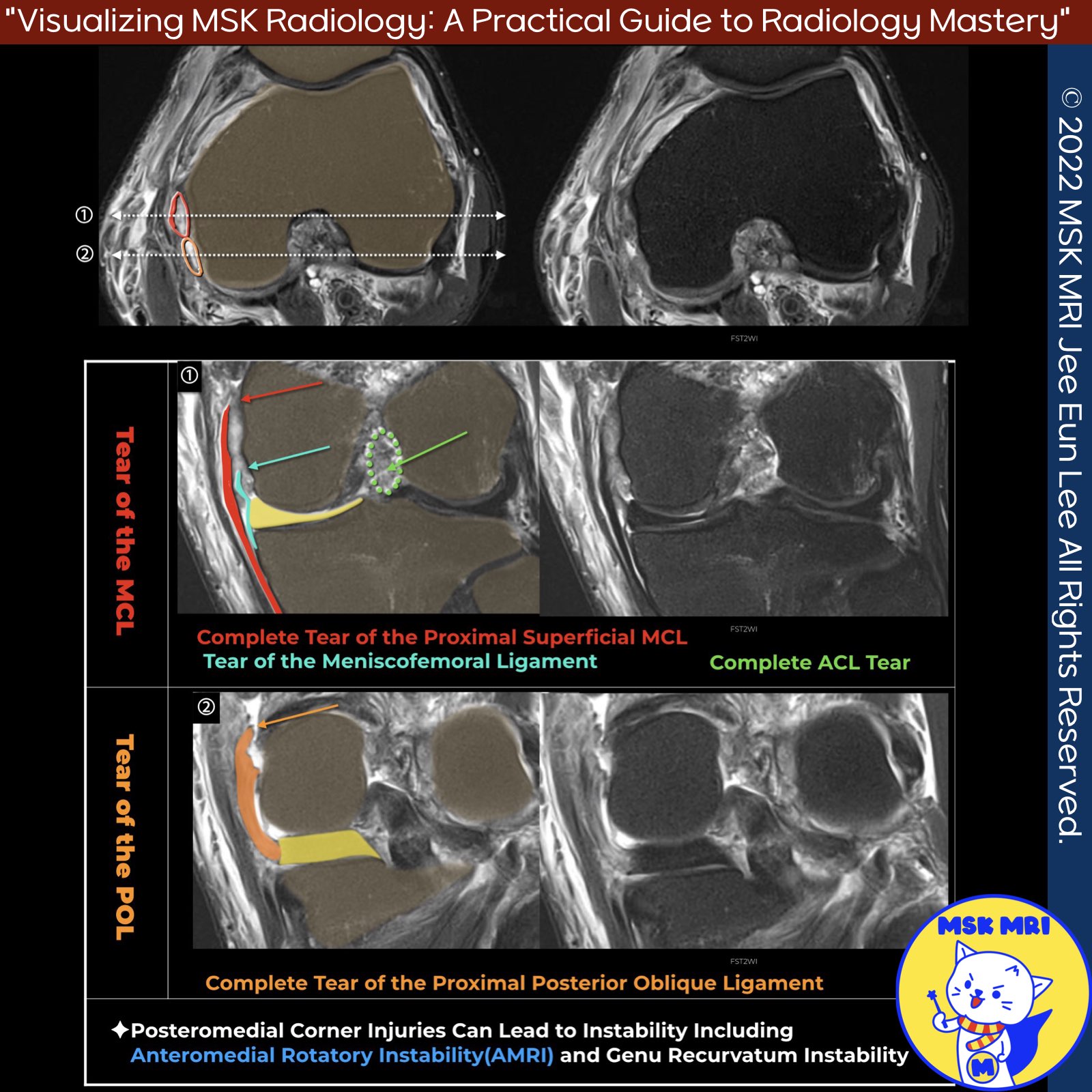
✅ Anatomy of the Posteromedial Corner
- Located between the superficial medial collateral ligament and posterior cruciate ligament
- Consists of 5 main structures: semimembranosus tendon, oblique popliteal ligament, posterior oblique ligament, posteromedial capsule, posterior horn of medial meniscus
✅ Biomechanical Importance
- Primary stabilizer against valgus laxity
- Secondary stabilizer against anterior tibial translation and external rotation
- Injuries can lead to anteromedial rotatory instability
📌 The Posterior Oblique Ligament
- Most frequently injured structure of the PMC
- Unlike the MCL, it controls rotational instability and valgus forces in knee extension
- Unrepaired injuries can cause chronic instability, early arthritis, reconstruction failure
✅ Clinical Significance
- PMC injuries, especially of the POL, must be identified
- Failure to treat can compromise ACL/ligament reconstruction outcomes
- Unaddressed, can lead to persistent instability and prevent full recovery
Radiographics. 2015 Jul-Aug;35(4):1123-37
Skeletal Radiol. 2022 May;51(5):1063-1071
J Am Acad Orthop Surg. 2017 Nov;25(11):752-761
Semin Musculoskelet Radiol. 2016 Feb;20(1):12-25.
"Visualizing MSK Radiology: A Practical Guide to Radiology Mastery"
© 2022 MSK MRI Jee Eun Lee All Rights Reserved.
No unauthorized reproduction, redistribution, or use for AI training.
#PMC, #POL, #Posteriorobliqueligament, #kneeMRI, #ACLtear, #MCLinjury, #AMRI, #kneeinstability


'✅ Knee MRI Mastery > Chap 3.Collateral Ligaments' 카테고리의 다른 글
| (Fig 3-A.30) Grade II Posterior Oblique Ligament Injury_ Part 1 (0) | 2024.05.11 |
|---|---|
| (Fig 3-A.29) Grade I Posterior Oblique Ligament Injury (0) | 2024.05.11 |
| (Fig 3-A.27) Posteromedial Corner MRI Anatomy, Posterior Oblique Ligament (0) | 2024.05.10 |
| (Fig 3-A.26) Three arms of the posterior oblique ligament, anatomy (0) | 2024.05.10 |
| (Fig 3-A.25) Reverse Segond Fracture (0) | 2024.05.09 |