Click the link to purchase on Amazon 🎉📚
==============================================
🎥 Check Out All Videos at Once! 📺
👉 Visit Visualizing MSK Blog to explore a wide range of videos! 🩻
https://visualizingmsk.blogspot.com/?view=magazine
📚 You can also find them on MSK MRI Blog and Naver Blog! 📖
https://www.instagram.com/msk_mri/
Click now to stay updated with the latest content! 🔍✨
==============================================
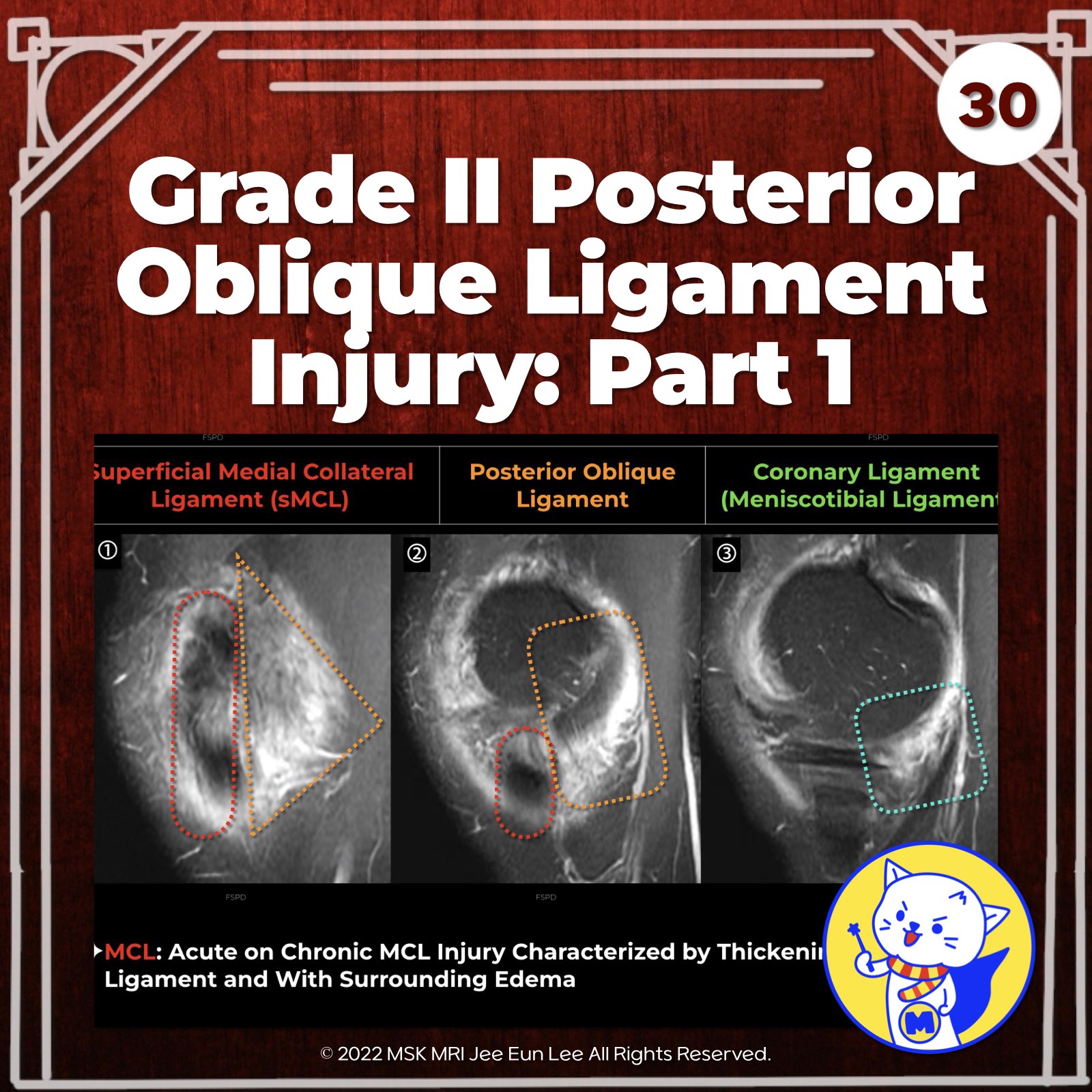
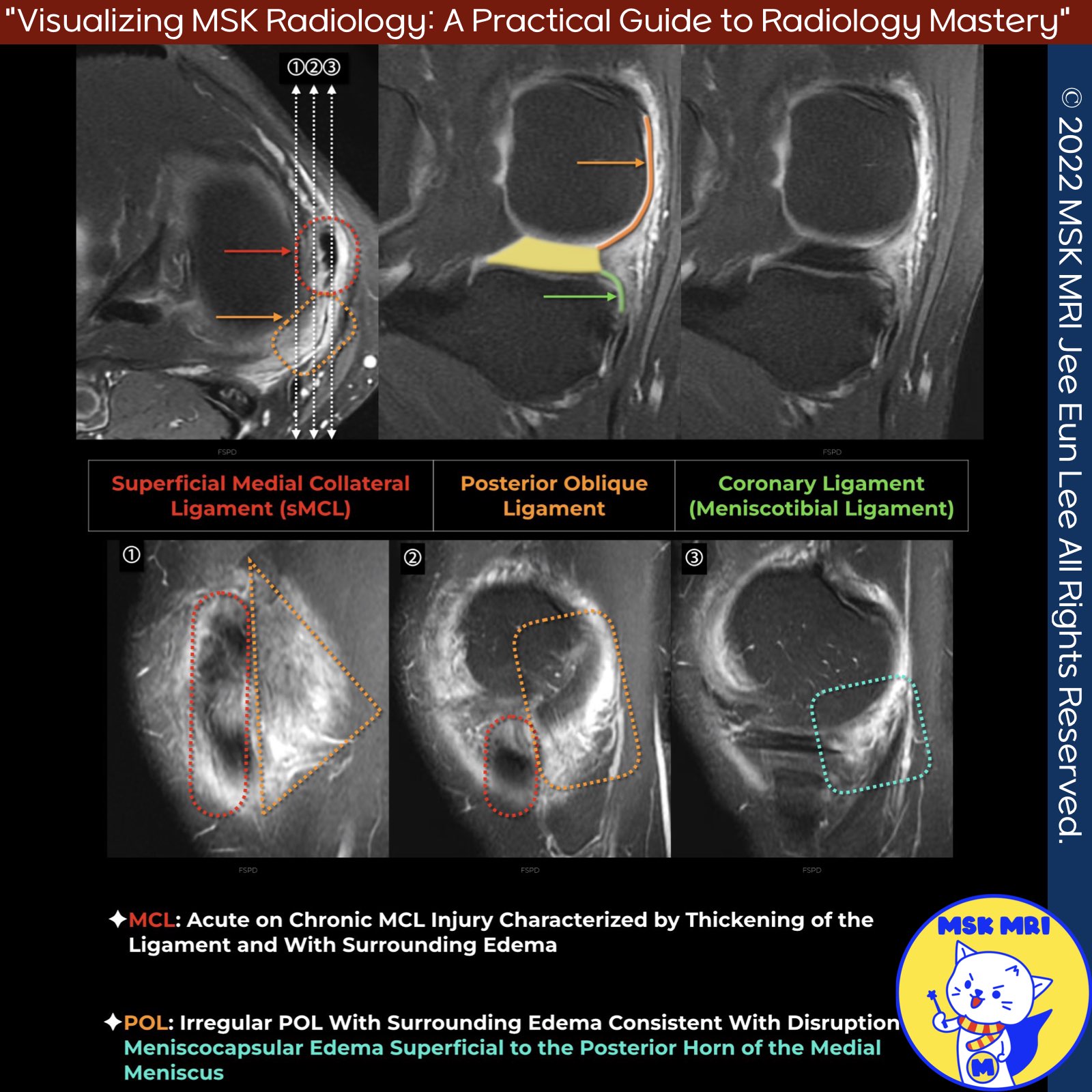
📌Posterior Oblique Ligament (POL)
- The POL is functionally distinct from the Medial Collateral Ligament (MCL) and is the most frequently injured structure of the Posteromedial Corner (PMC).
✅ Anatomy of the POL
- The POL originates proximally just behind and slightly inferior to the proximal origin of the superficial MCL (sMCL). It runs obliquely posteriorly and caudally to insert at the meniscus and the proximal tibia. Additionally, it connects to the semimembranosus, the oblique popliteal ligament (OPL), and the posteromedial capsule.
✅ Major Injury Patterns to the PMC
- POL Injury with Capsular Arm Involvement: Involves the capsular arm of the semimembranosus tendon (70%).
- POL Injury with Meniscal Detachment: Involves complete peripheral meniscal detachment (30%).
- Combined POL Injury: Includes disruption of the semimembranosus tendon and peripheral meniscal detachment (19%).
✅ Common Disruptions in Complex Knee Injuries
- Disruptions of the distal POL are frequent in complex knee injuries, notably in association with the oblique popliteal ligament, medial collateral ligament, and semimembranosus tendon tears.
Semin Musculoskelet Radiol. 2016 Feb;20(1):12-25
J Am Acad Orthop Surg. 2017 Nov;25(11):752-761
Skeletal Radiol. 2022 May;51(5):1063-1071
"Visualizing MSK Radiology: A Practical Guide to Radiology Mastery"
© 2022 MSK MRI Jee Eun Lee All Rights Reserved.
No unauthorized reproduction, redistribution, or use for AI training.
#PMC, #POL, #Posteriorobliqueligament, #kneeMRI, #ACLtear, #MCLinjury, #AMRI, #kneeinstability


'✅ Knee MRI Mastery > Chap 3.Collateral Ligaments' 카테고리의 다른 글
| (Fig 3-A.32) Grade III Posterior Oblique Ligament Injury (0) | 2024.05.11 |
|---|---|
| (Fig 3-A.31) Grade II Posterior Oblique Ligament Injury_ Part 2 (0) | 2024.05.11 |
| (Fig 3-A.29) Grade I Posterior Oblique Ligament Injury (0) | 2024.05.11 |
| (Fig 3-A.28) Anteromedial Rotatory Instability (0) | 2024.05.10 |
| (Fig 3-A.27) Posteromedial Corner MRI Anatomy, Posterior Oblique Ligament (0) | 2024.05.10 |