Click the link to purchase on Amazon 🎉📚
==============================================
🎥 Check Out All Videos at Once! 📺
👉 Visit Visualizing MSK Blog to explore a wide range of videos! 🩻
https://visualizingmsk.blogspot.com/?view=magazine
📚 You can also find them on MSK MRI Blog and Naver Blog! 📖
https://www.instagram.com/msk_mri/
Click now to stay updated with the latest content! 🔍✨
==============================================
출처: https://mskmri.tistory.com/1056 [MSK MRI:티스토리]
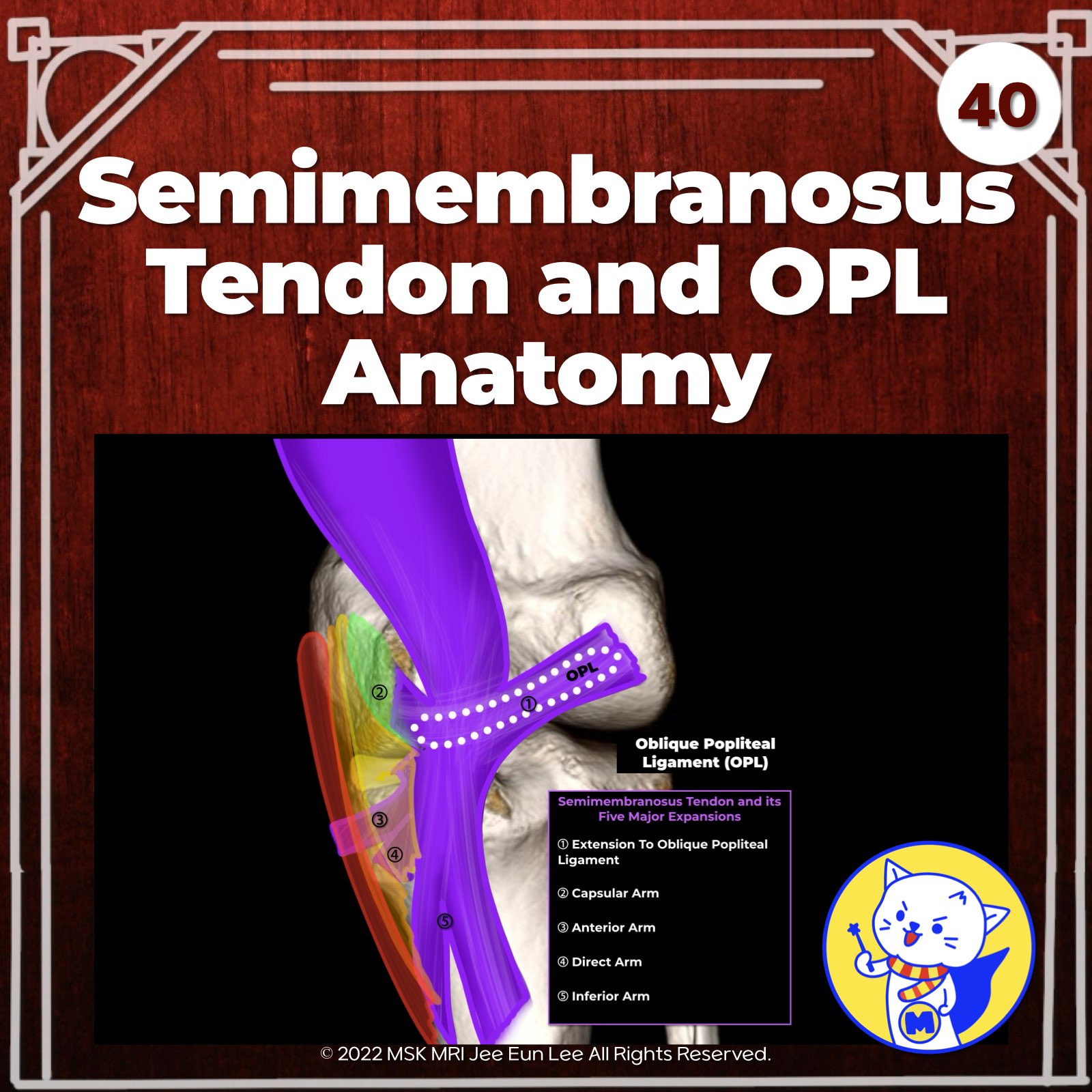
📌 Oblique Popliteal Ligament
- The oblique popliteal ligament (OPL) originates medially from the merging of the semimembranosus's lateral expansion and the capsular arm of the popliteus ligament (POL).
- Laterally, it attaches to the fabella, the meniscofemoral portion of the posterolateral joint capsule, and the plantaris muscle.
- The OPL, a lateral extension of the semimembranosus tendon, surrounds the posteromedial joint capsule and is the largest structure in the posterior knee, forming a fascial sheet approximately 5 cm long and 1–1.5 cm wide.
Radiographics. 2015 Jul-Aug;35(4):1123-37
Radiol Clin North Am. 2013 May;51(3):413-32
Skeletal Radiol. 2014 Jun;43(6):781-91.
"Visualizing MSK Radiology: A Practical Guide to Radiology Mastery"
© 2022 MSK MRI Jee Eun Lee All Rights Reserved.
No unauthorized reproduction, redistribution, or use for AI training.
#semimembranosus, #kneeMRI, #Kneeanatomy, #semimembranosustendon, #Fabella, #OPL, #POL, #jointcapsule


'✅ Knee MRI Mastery > Chap 3.Collateral Ligaments' 카테고리의 다른 글
| (Fig 3-A.42) Oblique Popliteal Ligament Injury (0) | 2024.05.13 |
|---|---|
| (Fig 3-A.41) Oblique Popliteal Ligament MRI Anatomy (0) | 2024.05.13 |
| (Fig 3-A.39) Fat Accumulation in Semimembranosus Tendon (0) | 2024.05.12 |
| (Fig 3-A.38) Semimembranosus Tendinopathy: Part 2 (0) | 2024.05.12 |
| (Fig 3-A.37) Semimembranosus Tendinopathy: Part 1 (0) | 2024.05.12 |