Click the link to purchase on Amazon 🎉📚
==============================================
🎥 Check Out All Videos at Once! 📺
👉 Visit Visualizing MSK Blog to explore a wide range of videos! 🩻
https://visualizingmsk.blogspot.com/?view=magazine
📚 You can also find them on MSK MRI Blog and Naver Blog! 📖
https://www.instagram.com/msk_mri/
Click now to stay updated with the latest content! 🔍✨
==============================================
출처: https://mskmri.tistory.com/1056 [MSK MRI:티스토리]
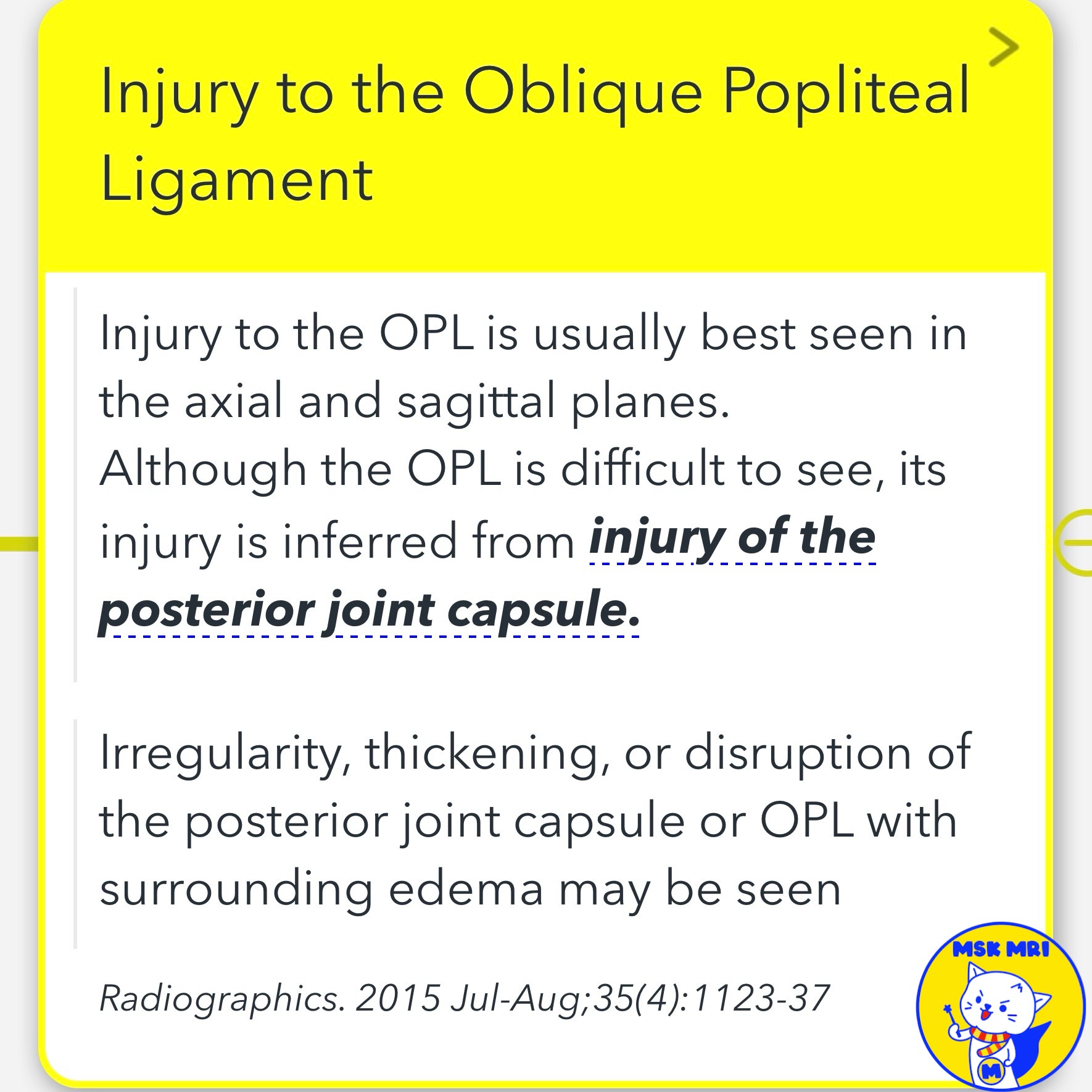
📌 Injury to the Oblique Popliteal Ligament
- Injury is usually best seen on axial and sagittal planes
- Difficult to visualize the intact ligament directly
- Inferred from injury to the posterior joint capsule
- Signs include irregularity, thickening, disruption of the posterior capsule/OPL and surrounding edema
- Edema and irregularity of the posteromedial joint capsule suggesting high-grade oblique popliteal ligament tear
- Disruption of the posterior joint capsule implying at least partial oblique popliteal ligament tear
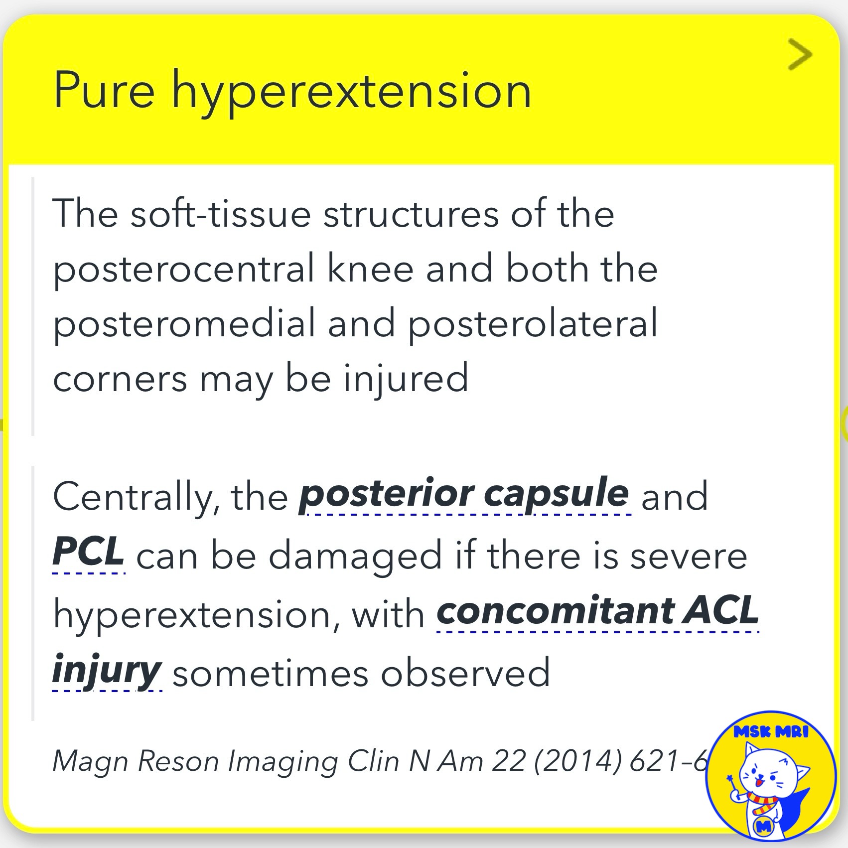
✅ Mechanism of Injury: Pure Hyperextension
- May injure soft tissues around the posterocentral knee
- Both posteromedial and posterolateral corners can be affected
- Central injuries can involve the posterior capsule and PCL with severe hyperextension
- Concomitant ACL injury sometimes observed
Radiographics. 2015 Jul-Aug;35(4):1123-37
Skeletal Radiol (2014) 43:239–242
Magn Reson Imaging Clin N Am 22 (2014) 621–648
"Visualizing MSK Radiology: A Practical Guide to Radiology Mastery"
© 2022 MSK MRI Jee Eun Lee All Rights Reserved.
No unauthorized reproduction, redistribution, or use for AI training.
#jointcapsuleinjury, #kneeMRI, #Kneeanatomy, #Hyperextensioninjury, #PCLavulsionfracture, #PCLinjury, #OPL, #jointcapsule


'✅ Knee MRI Mastery > Chap 3.Collateral Ligaments' 카테고리의 다른 글
| (Fig 3-A.44) O’Donoghue’s Pentad Lesion (0) | 2024.05.13 |
|---|---|
| (Fig 3-A.43) O'Donoghue’s Triad Lesion (0) | 2024.05.13 |
| (Fig 3-A.41) Oblique Popliteal Ligament MRI Anatomy (0) | 2024.05.13 |
| (Fig 3-A.40) Semimembranosus Tendon and OPL Anatomy (0) | 2024.05.13 |
| (Fig 3-A.39) Fat Accumulation in Semimembranosus Tendon (0) | 2024.05.12 |