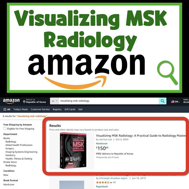
Click the link to purchase on Amazon 🎉📚
==============================================
🎥 Check Out All Videos at Once! 📺
👉 Visit Visualizing MSK Blog to explore a wide range of videos! 🩻
https://visualizingmsk.blogspot.com/?view=magazine
📚 You can also find them on MSK MRI Blog and Naver Blog! 📖
https://www.instagram.com/msk_mri/
Click now to stay updated with the latest content! 🔍✨
==============================================
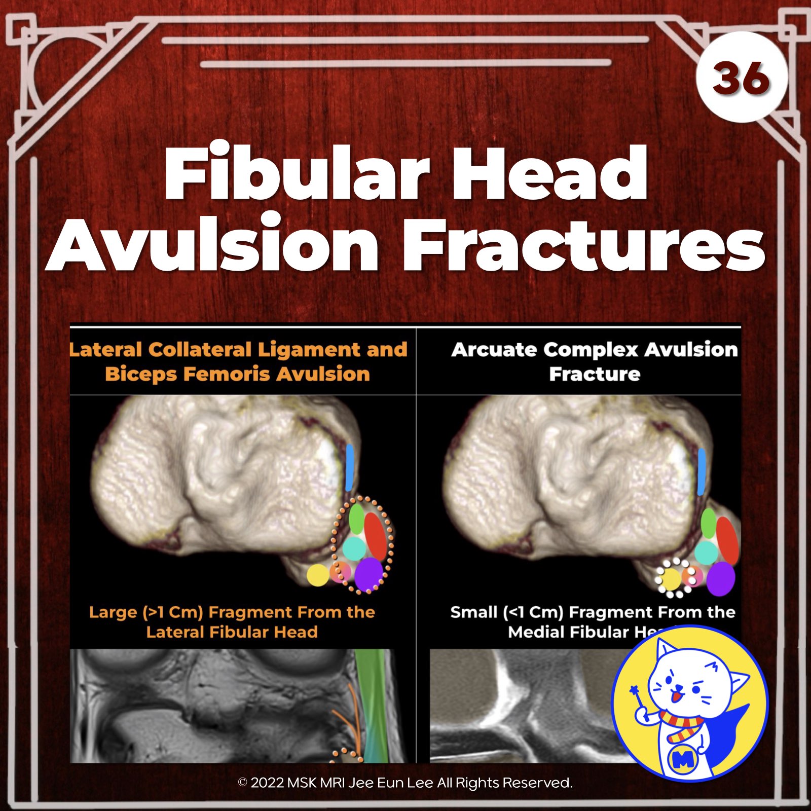
📌 Arcuate Fractures and Posterior Lateral Corner (PLC) Injuries
Introduction Certain osseous injuries, known as arcuate fractures or the arcuate sign, provide diagnostic clues for an underlying PLC injury, alerting radiologists to inspect this region closely. (Rosenberg et al., 2016)
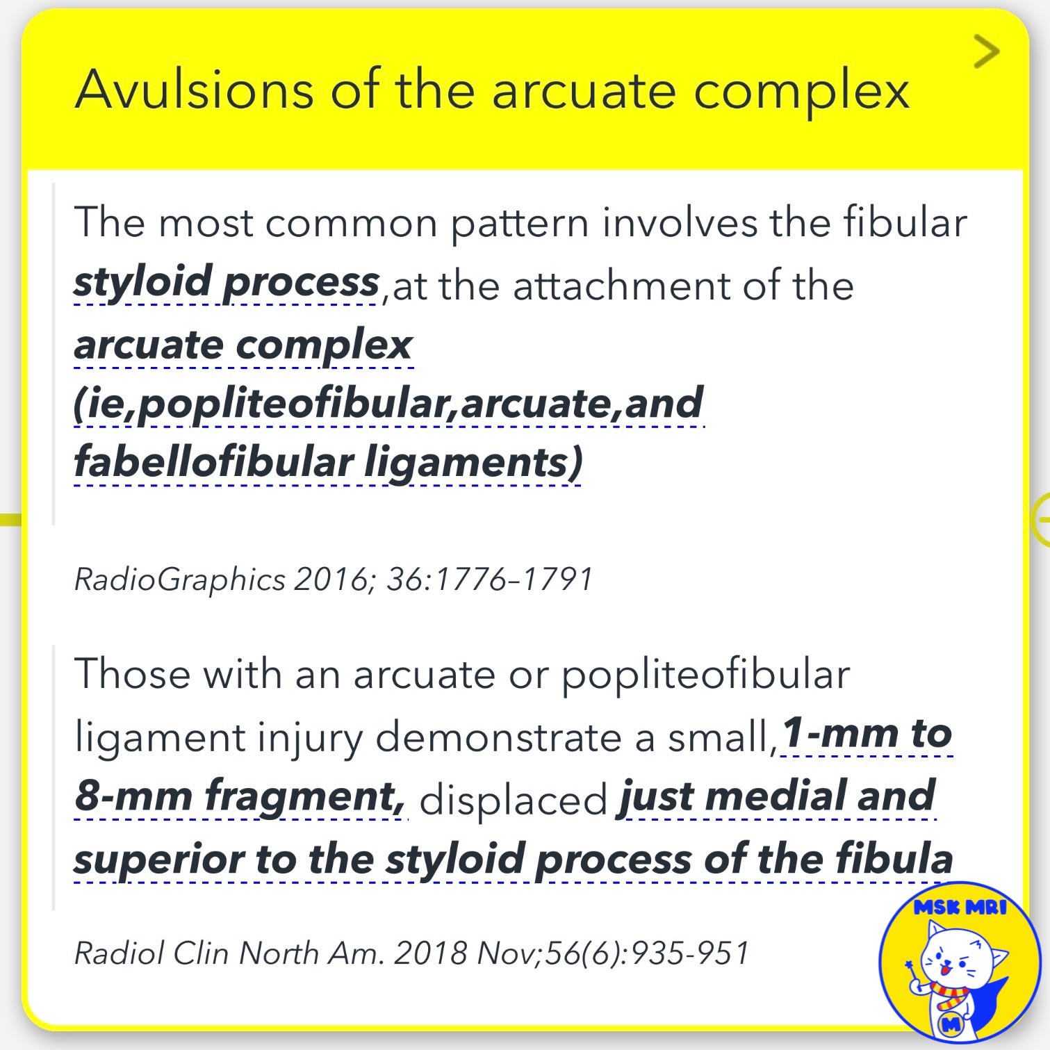
✅ Avulsions of the Arcuate Complex
- The most common pattern involves the fibular styloid process at the attachment of the arcuate complex (popliteofibular, arcuate, and fabellofibular ligaments). (Rosenberg et al., 2016)
- These injuries demonstrate a small, 1-mm to 8-mm fragment displaced just medial and superior to the fibula's styloid process. (Bosmans et al., 2018)
✅ Avulsions of the Lateral Collateral Ligament (LCL) and Biceps Femoris Tendon
- Fractures resulting from avulsions of the LCL and biceps femoris tendon are larger and involve the lateral margin of the fibular head. (Rosenberg et al., 2016)
- These fractures measure 1.5 to 2.5 cm in size. (Bosmans et al., 2018)
✅ Differentiating Arcuate Complex and LCL/Biceps Femoris Tendon Injuries
- The presence and size of these avulsion fractures can help differentiate between injuries to the arcuate complex and injuries to the LCL and biceps femoris tendon, which are components of the PLC. (Rosenberg et al., 2016; Bosmans et al., 2018)
RadioGraphics 2016; 36:1776–1791
Radiol Clin North Am. 2018 Nov;56(6):935-951
"Visualizing MSK Radiology: A Practical Guide to Radiology Mastery"
© 2022 MSK MRI Jee Eun Lee All Rights Reserved.
No unauthorized reproduction, redistribution, or use for AI training.
#ArcuateFractures, #PosteriorLateralCornerInjury, #AvulsionFractures, #FibulaHead, #ArcuateComplex, #LateralCollateralLigament, #BicepsFemorisTendon, #AvulsionFragments, #RadiologicFindings, #KneeTraumaRadiology


'✅ Knee MRI Mastery > Chap 3.Collateral Ligaments' 카테고리의 다른 글
| (Fig 3-B.38, 39, 42) Posterolateral Corner Reconstruction (0) | 2024.05.24 |
|---|---|
| (Fig 3-B.37) Anterior Medial Tibial Plateau Rim Fracture (0) | 2024.05.24 |
| (Fig 3-B.35) Fibula Head Attachments Anatomy (0) | 2024.05.24 |
| (Fig 3-B.34) Iliotibial Band Friction Syndrome (0) | 2024.05.24 |
| (Fig 3-B.33) Iliotibial Band Injury from Acute Trauma (0) | 2024.05.23 |