https://youtu.be/vzxclU_34OI?si=B88NykUJHSELzyAC
Click the link to purchase on Amazon 🎉📚
==============================================
🎥 Check Out All Videos at Once! 📺
👉 Visit Visualizing MSK Blog to explore a wide range of videos! 🩻
https://visualizingmsk.blogspot.com/?view=magazine
📚 You can also find them on MSK MRI Blog and Naver Blog! 📖
https://www.instagram.com/msk_mri/
Click now to stay updated with the latest content! 🔍✨
==============================================
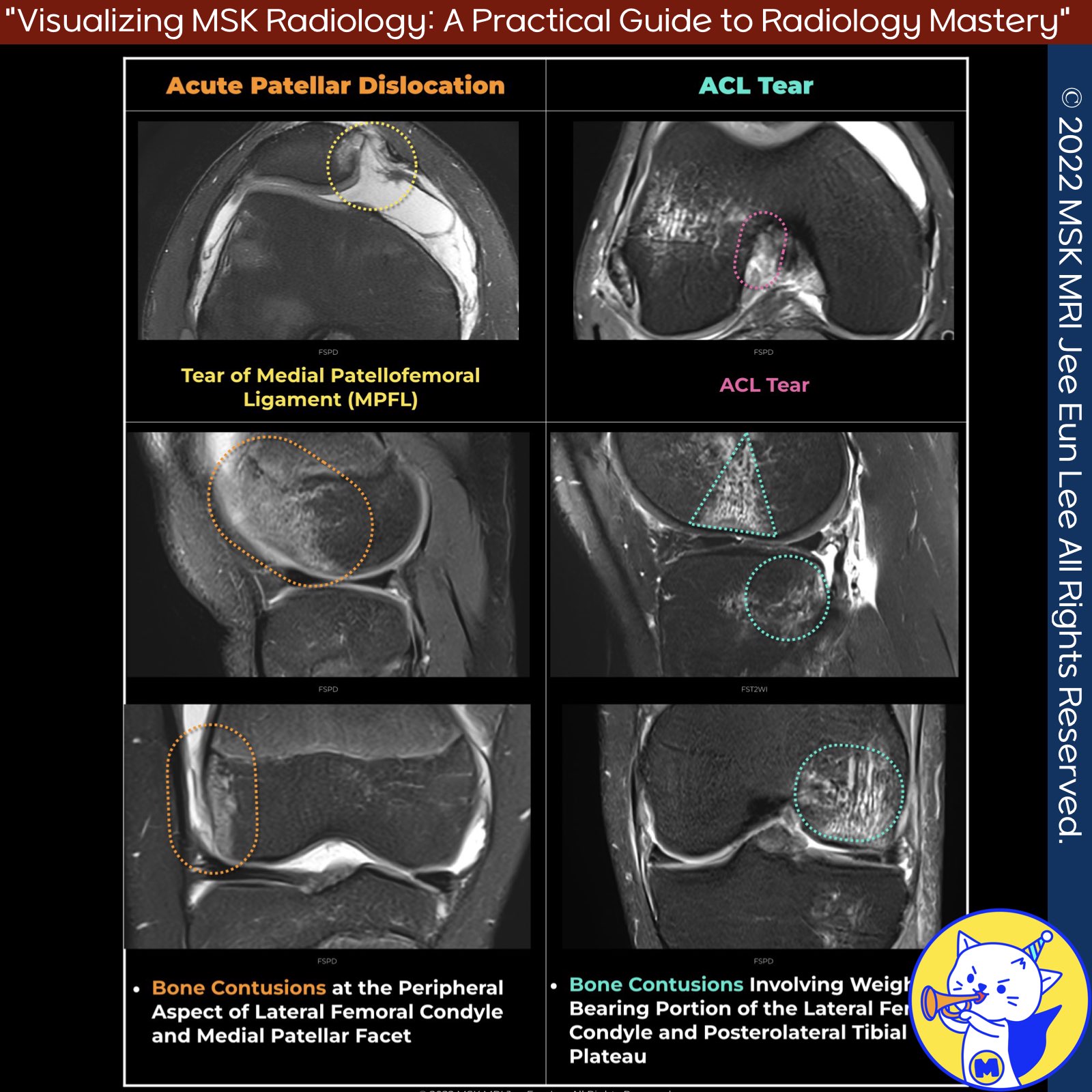
📌 Differentiating Patellofemoral Instability from ACL Tears Based on Bone Bruise Patterns and Osteochondral Injury
1️⃣ Patellofemoral Instability
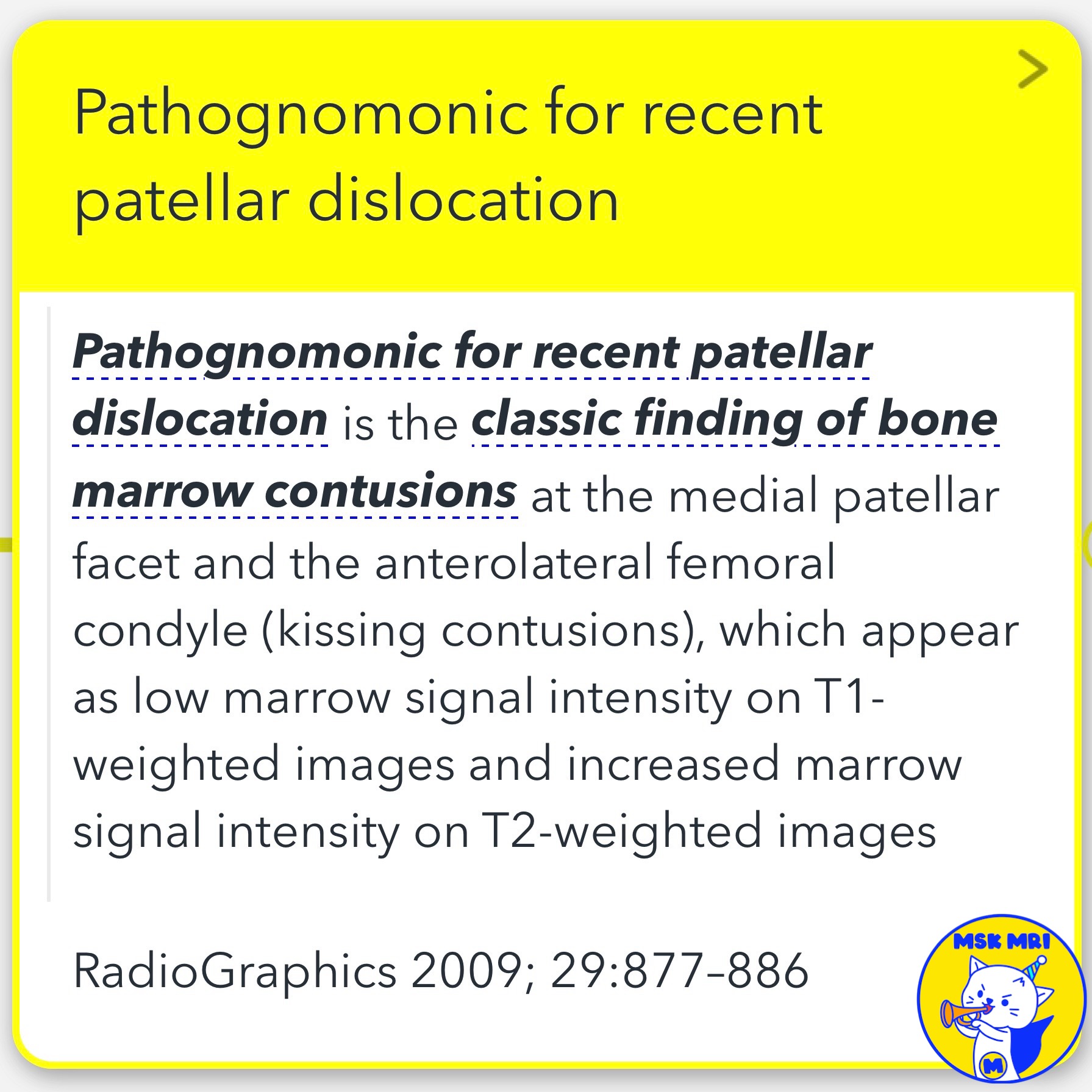
✅ Pathognomonic Signs
- Recent patellar dislocation has classic imaging findings of bone marrow contusions at the medial patellar facet and the anterolateral femoral condyle. These are also known as "kissing contusions."
- On MRI, these contusions appear as low marrow signal intensity on T1-weighted images and increased marrow signal intensity on T2-weighted images. This pattern is highly indicative of a recent patellar dislocation.
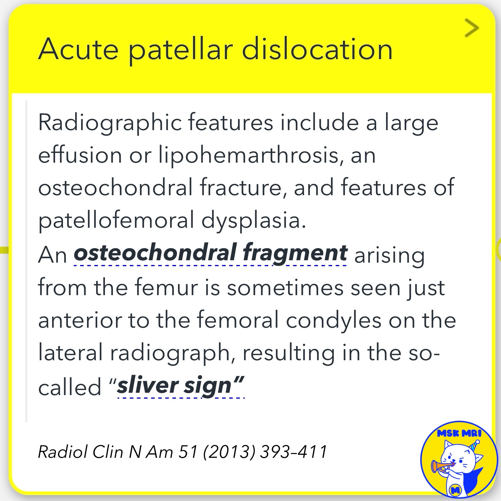
✅ Radiographic Features
- We often see a large effusion or lipohemarthrosis.
- An osteochondral fracture may be present, along with signs of patellofemoral dysplasia.
- Occasionally, an osteochondral fragment from the femur can be seen just anterior to the femoral condyles on a lateral radiograph, known as the “sliver sign.”
2️⃣ ACL Tears
✅ Bone Bruise Patterns
- ACL injuries commonly involve osseous injuries at the anterior aspect of the lateral femoral condyle and the posterior aspect of the lateral tibial plateau.
- Lateral compartment osseous contusions are most specifically found at the posterolateral tibial plateau. Less commonly, these contusions may also occur at the posterior aspect of the medial tibial plateau and medial femoral condyle.
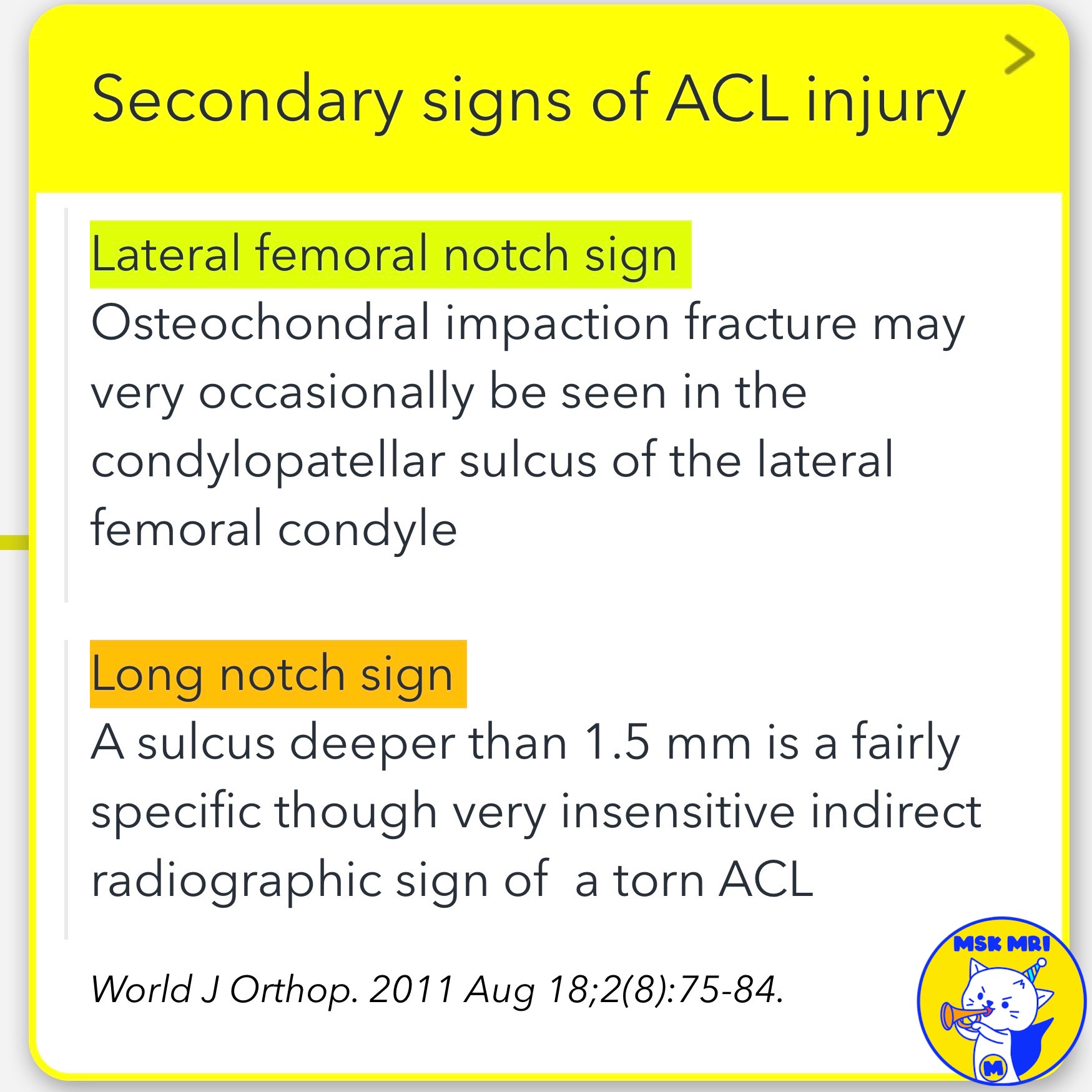
✅ Secondary Signs
- The lateral femoral notch sign is another indicator, where an osteochondral impaction fracture may be seen in the condylopatellar sulcus of the lateral femoral condyle.
- The “long notch sign” refers to a sulcus deeper than 1.5 mm, which is a specific, though not very sensitive, indirect radiographic sign of a torn ACL.
References:
- Radiographics 2009; 29:877–886.
- Radiol Clin N Am 51 (2013) 393–411.
- Magn Reson Imaging Clin N Am 30 (2022) 261–275.
- World J Orthop. 2011 Aug 18;2(8):75-84.
"Visualizing MSK Radiology: A Practical Guide to Radiology Mastery"
© 2022 MSK MRI Jee Eun Lee All Rights Reserved.
No unauthorized reproduction, redistribution, or use for AI training.
#PatellofemoralInstability, #ACLTear, #BoneBruise, #OsteochondralInjury, #KneeInjury, #MRI, #MedicalImaging, #Orthopedics, #SportsMedicine


'✅ Knee MRI Mastery > Chap 4A. Patelloefemoral joint' 카테고리의 다른 글
| (Fig 4-A.25) Chondral Delamination and Flap in Patellar Dislocation (0) | 2024.06.03 |
|---|---|
| (Fig 4-A.24) Osteochondral Injury in Patellar Dislocation (0) | 2024.06.03 |
| (Fig 4-A.22) Recent Patellar Dislocation: Key Findings (0) | 2024.06.02 |
| (Fig 4-A.21) Excessive Femoral Anteversion (0) | 2024.06.02 |
| (Fig 4-A.20) Tibial Tubercle Lateralization (0) | 2024.06.02 |