본문 바로가기

Knee MRI62

[Knee-04] Anatomy of layers and components, Posterolateral corner 01 
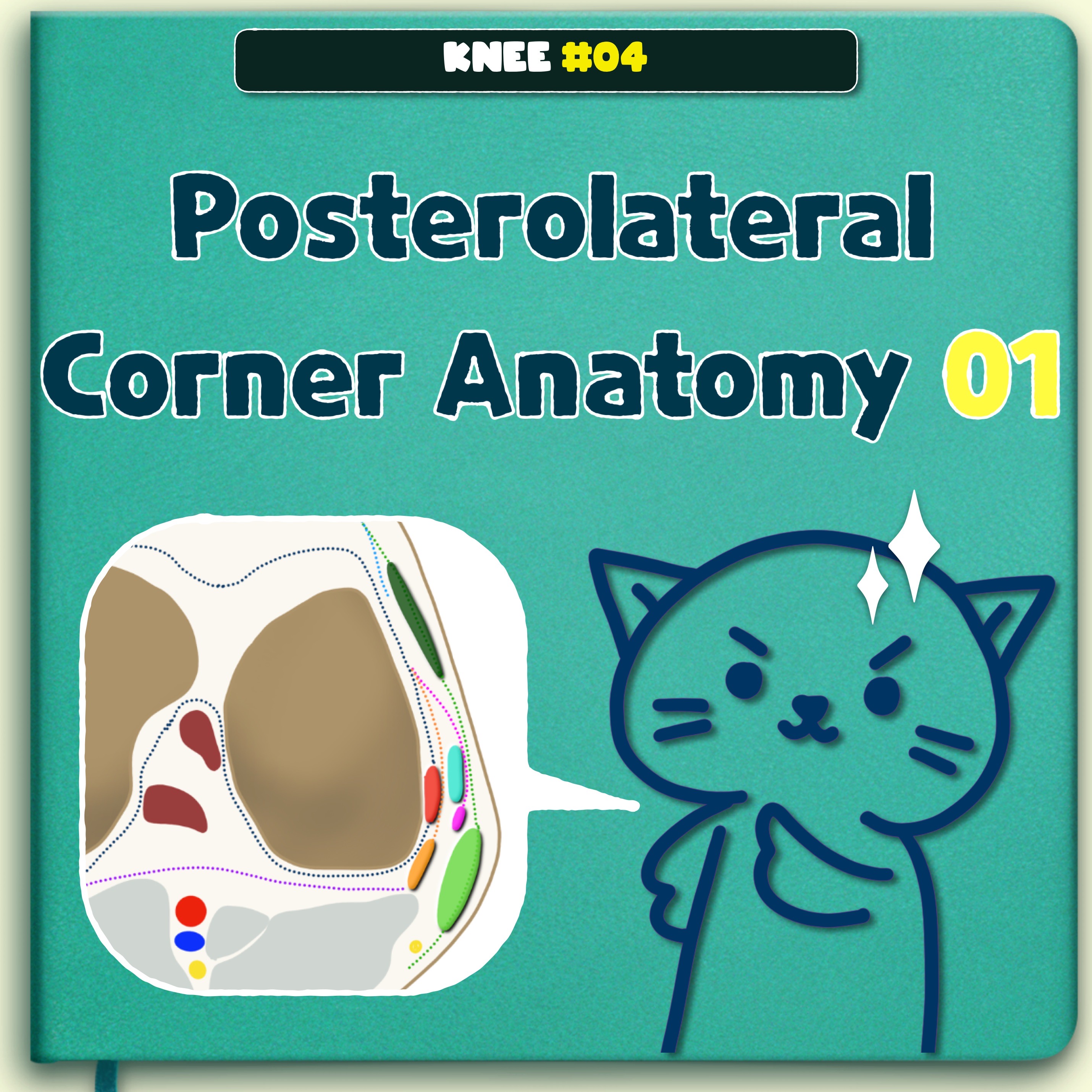
https://youtu.be/lMx9cjkzNZw Three structural layers Layer 1:most superficial Fascia lata lliotibial tract with its anterior expansion Superficial portion of biceps femoris with its posterior expansion ​ Layer 2 Quadriceps retinaculum anteriorly Two patellofemoral ligaments or retinacula posteriorly Lavers 1 and 2 merges at lateral aspect of the patella ​ Layer 3:deepest layer The lateral joint .. 2022. 12. 25.
[Knee-03] Tear of Popliteus tendon, Posterolateral corner (PLC) injury https://youtu.be/XVRwYJa4f2M 1. Tear of the popliteus tendon A. Posterolateral (arcuate) complex LCL Popliteus tendon Lateral head of gastrocnemius muscle Arcuate ligament Popliteofibular ligament Fabellofibular ligament Stoller's Orthopaedics and Sports Medicine: The Knee B. Posterolateral corner (PLC) injury Injury to a component of the lateral collateral ligament complex in association with a.. 2022. 12. 24.
[Knee-02] Popliteus tendon anatomy, Posterolateral attachment https://youtu.be/pkdL_HwgPi0 Popliteus The popliteus musculotendinous complex—consisting of the popliteus tendon, popliteus muscle, and PFL The tendon, which is intracapsular, but extra-articular and extrasynovial, ascends around the posterolateral aspect of the knee The tendinous attachment lies anteroinferior to the proximal attachment of the lateral collateral ligament on the lateral epicondy.. 2022. 12. 24.
[HYPERLINK] Anterolateral Ligament, ACL tear, Anterolateral reconstruction, Segond fracture (SITE MAP) Anterolateral Ligament, ACL tear, Anterolateral reconstruction, Segond fracture ​ [ALL_01] #01 Anatomy, Anterolateral Ligament, Knee MRI ​ [ALL_02] #02 Anatomy, Anterolateral Ligament, Knee MRI, ​ [ALL_03] #01 Injury to the Anterolateral Ligament, MRI, US, Knee, Segond fracture [ALL_04] #02 Injury to the Anterolateral Ligament, MRI, US, Knee, Segond fracture ​ [ALL_05] Anterolateral reconstructi.. 2022. 5. 14.
[HYPERLINK] Meniscal tear, horizontal, longitudinal, flap, root tear, Bucket han Meniscal tear, horizontal, longitudinal, flap, root tear, Bucket handle, Meniscectomy, Meniscal repair, meniscus allograft transplant ​ Meniscal Tear [Meniscal Tear_01] Horizontal tear, knee MRI, Horizontal cleavage tear [Meniscal Tear_02] Longitudinal vertical tear-1, knee MRI, Wrisberg Rip, Popliteomeniscal fascicles [Meniscal Tear_03] Longitudinal vertical tear-2, knee MRI, Wrisberg Rip, Popl.. 2022. 5. 14.
[HYPERLINK] Meniscus Anatomy, root, Discoid meniscus, Wrisberg ligament, Poplite Meniscus Anatomy, root, Discoid meniscus, Wrisberg ligament, Popliteomeniscal fascicles, Variants and Pitfalls ​ Anatomy ​ [Meniscus Anatomy_01] Knee MRI, Anatomy, anterior root, posterior root, medial lateral meniscus [Meniscus Anatomy_02] Popliteomeniscal fascicles (anteroinferior, posterosuperior ) [Meniscus Anatomy_03] Humphry and Wrisberg ligaments, meniscotibial ligament, knee MRI [Meniscu.. 2022. 5. 14.
[HYPERLINK] Anterior Cruciate Ligament Repair and Its Complications (SITE MAP) Anterior Cruciate Ligament Repair and Its Complications ​ 전방십자인대 재건술 -1 (introduction) /무릎 MRI ​ 전방십자인대 재건술 -2 (수술법 및 graft MRI 소견) /무릎 MRI 전방십자인대 재건술 -3 (터널 위치) /무릎 MRI​ 전방십자인대 재건술 -4 (수술실패-충돌) /무릎 MRI ​ 전방십자인대 재건술 -5 (수술실패-관절섬유증, 유리체) /무릎 MRI 전방십자인대 재건술 -6 (수술실패-불안정성, 이식건 파열) /무릎 MRI 전방십자인대 재건술 -7 (수술실패-고정관련 실패) /무릎 MRI ​ 전방십자인대 재건술 -8 (수술실패-터널확장, 자가건 채취부위) /무릎 MRI​ 전방십자인대 재건술 -9 (수술실패-장경인대 마찰.. 2022. 5. 14.
[HYPERLINK] Knee MRI checklist, meniscus, tear, ACL, PCL, MCL, LCL, Extensor mec Knee MRI checklist ​ [Knee_01] MRI check list, meniscus, anatomy, root, horizontal tear, vertical tear, meniscus tear [Knee_02] Meniscus tear, radial tear, root tear, flap, parrot beak , Bucket handle tear, KNEE MRI [Knee_03] meniscectomy, meniscus repair, Transtibial pullout repair, transplantation, KNEE MRI [Knee_04] ACL, PCL, Tear, Reconstruction, femoral tunnel, tibial tunnel, impingement, g.. 2022. 5. 14.
[Tear_30] Ramp lesion_Summary_meniscocapaular tear, Grief, Thaunat classificatio Meniscal ramp lesions can be defined as longitudinal vertical and/or oblique peripheral tears in the posterior horn of medial meniscus, in a mediolateral direction of less than 2.0 cm, that may lead to meniscocapsular or meniscotibial disruption with a concomitant ACL injury. There are few classifications regarding meniscal ramp lesions. Thaunat et al. approached a more comprehensive classificat.. 2021. 10. 23.
[Tear_29] Ramp lesion_Pitfalls, Type 4A vs 4B lesion, meniscocapsular separation The subtype 4A is a complete longitudinal vertical tear of the red-red zone of the meniscus, The subtype 4B involves a complete tear of the junction itself where the meniscocapsular and meniscotibial fibers attach to the posterior horn. A very peripheral meniscal tear(ramp type 4A lesion) can mimic ramp type 4B lesion. (Type 4B ramp lesion) There are edema and irregularity of the meniscocapsular.. 2021. 10. 23.
[Tear_28] Ramp lesion_Pitfalls, perimeniscal fluid, edema of posterior capsule https://youtu.be/HLYXPEdQ9Mc The perimeniscal fluid, edema of the posterior capsule, or even normal structures can be mistaken for a ramp lesion. Let's look at the normal meniscocapsular junction and find out the difference between these lesions and ramp lesions. The meniscocapsular junction of the posterior horn of medial meniscus is composed of the meniscocapsular ligament superiorly and the m.. 2021. 10. 23.
[Tear_27] Ramp lesion_Type 5: peripheral posterior horn meniscal double tear Finally, look at the type 5 ramp lesion. In this tear pattern, there are two separate tears within the red- red zone of the meniscus. So this is called a double tear. Type 5 ramp lesions are similar to type 4a. The subtype 4A is a complete longitudinal vertical tear of the red-red zone of the meniscus, with the meniscocapsular and meniscotibial ligaments intact but connected to a portion of the .. 2021. 10. 22.
[Tear_26] Ramp lesion_Type 4B: complete meniscocapsular junction tear Type 4 lesions involve a complete longitudinal vertical tear in the posterior horn of the meniscus, leading to meniscotibial ligament instability. There are two different subtypes of type 4 lesions in order to account for the instability pattern. The subtype 4A is a complete longitudinal vertical tear of the red-red zone of the meniscus, The second pattern (subtype 4B) involves a complete tear o.. 2021. 10. 22.
[Tear_25] Ramp lesion_Type 4A: complete peripheral posterior horn meniscal tear Type 4 lesions as described by Thaunat et al. involve a complete longitudinal vertical tear in the posterior horn of the meniscus, leading to meniscotibial ligament instability as seen with type 3 lesions. To better account for the instability pattern seen with complete tears, Grief et al proposed further two subtypes of this type 4 ramp lesion. The subtype 4A is a complete longitudinal vertical.. 2021. 10. 21.
[Tear_24] Ramp lesion_Meniscocapsular Separations_02 Longitudinal tears of the posterior horn or meniscocapsular junction can propagate anteriorly into the meniscal periphery at the junction of body and posterior horn. There is only a partial separation of the meniscocapsular junction occurring as a continuation of a meniscal tear, producing a corner lesion. In this case, the sagittal image demonstrates the type 3A Ramp lesion, but this tear propa.. 2021. 10. 21.
[Tear_23] Ramp lesion_Meniscocapsular Separations_01 Meniscocapsular Separation is the tear of the supporting meniscotibial (coronary) or meniscofemoral ligaments typically at attachment to meniscus. Occasionally, there is only a partial separation of the meniscocapsular junction occurring as a continuation of a meniscal tear, producing a corner lesion. Left image: This meniscocapsular separation is type 3A ramp lesion which is partial peripheral .. 2021. 10. 21.
[Tear_22] Ramp lesion_Type 3B: meniscotibial ligament tear https://youtu.be/wGrw05qFUh0 There are two different subtypes of type 3 lesions in order to account for the meniscotibial ligament instability. Type 3A represents a vertical peripheral tear of the inferior margin of the posterior horn containing the attachment of the meniscotibial ligament. The other subtype (type 3B) is a tear of the meniscotibial ligament itself from its attachment to the post.. 2021. 10. 20.
[Tear_21] Ramp lesion_Type 3A: partial inferior peripheral posterior horn menisc Type 3 lesions, also known as “hidden lesions,” occur in the most inferior portion of the posterior horn and are associated with meniscotibial ligament instability. They are not visible with the transnotch approach, but they may be suspected in case of mobility at probing. There are two different subtypes of type 3 lesions in order to account for the meniscotibial ligament instability. Type 3A r.. 2021. 10. 20.
[Tear_20] Ramp lesion_Type 2: partial superior peripheral posterior meniscal hor Ramp lesion type 2 is a peripheral partial thickness tear involving the superior margin of the posterior horn, where the torn fragment contains the intact meniscocapsular attachment to the posterior horn. Linear vertical fluid signal reaching the superior articular surface of the posterior horn of the medial meniscus red-red zone. (Partial thickness superior vertical tear through the posterior h.. 2021. 10. 19.
[Tear_19] Ramp lesion_Type 1: meniscocapsular ligament tear Type 1 Ramp lesion is an isolated posterior superior meniscocapsular tear. Meniscocapsular tears are located peripherally and involve the synovium, leading to posterior meniscocapsular separation from the posterior horn of the medial meniscus. The meniscotibial ligament and the posterior horn itself remain intact. This tear pattern is commonly located in the posteromedial “blind spot” and is par.. 2021. 10. 19.
[Tear_18] Ramp lesion_Normal anatomy and mean length_02 The posterior meniscotibial ligament attachment to the PHMM had a mean length of 14.0 ± 5.4 mm at its insertion on the posterior tibia. The posterior meniscocapsular attachment to the PHMM had a mean length of 20.2 ± 6.0 mm. The dMCL had a broad, firm attachment to the midbody of the medial meniscus. The dMCL meniscofemoral attachment blended with the POL meniscofemoral attachment posteriorly an.. 2021. 10. 19.