Click the link to purchase on Amazon 🎉📚
==============================================
🎥 Check Out All Videos at Once! 📺
👉 Visit Visualizing MSK Blog to explore a wide range of videos! 🩻
https://visualizingmsk.blogspot.com/?view=magazine
📚 You can also find them on MSK MRI Blog and Naver Blog! 📖
https://www.instagram.com/msk_mri/
Click now to stay updated with the latest content! 🔍✨
==============================================
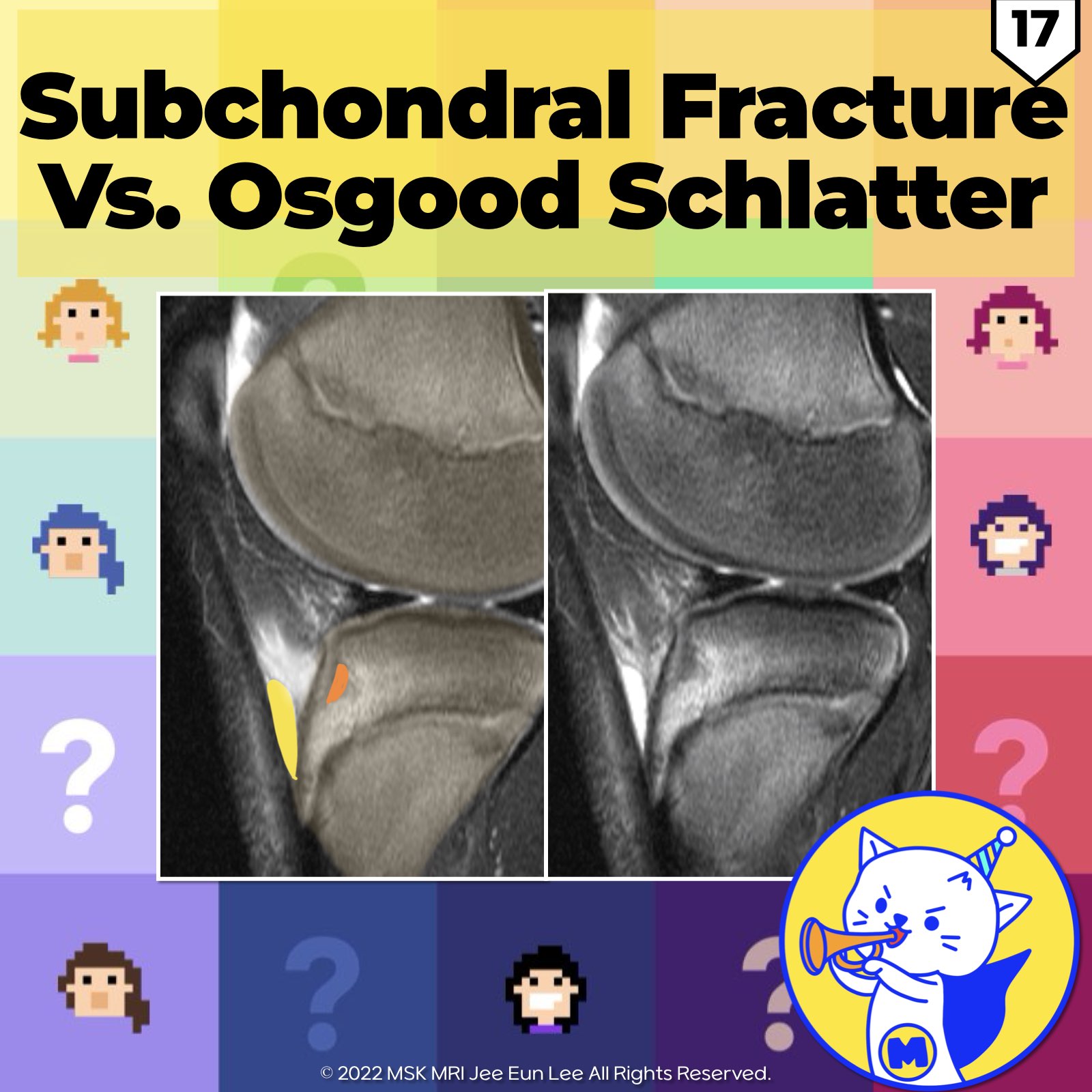
📌 Cartilaginous Stage of Osgood Schlatter Disease
✅ During the cartilaginous stage of tuberosity development in Osgood Schlatter disease
- Tuberosity marrow edema
- Subtle avulsive injuries to the secondary ossification center, often presenting as transverse clefts in the damaged ossifying cartilage, which may not be apparent on initial radiographs.
- Tendon thickening
- Prepatellar edema
- Deep infrapatellar bursitis
Reference: RadioGraphics 2018; 38:2069–2101
📌 Subchondral Fracture
✅ A subchondral fracture appears on MRI as:
- Linear areas of low T1 and T2 signal intensity, typically in regions of bone contusion
- These do not result in cortical disruption or deformity
- Seen as linear hypointensities beneath the cartilage without causing contour deformities or visible involvement of the articular surface.
References: AJR 2019; 213:963–982; RadioGraphics 2018; 38:1478–1495
"Visualizing MSK Radiology: A Practical Guide to Radiology Mastery"
© 2022 MSK MRI Jee Eun Lee All Rights Reserved.
No unauthorized reproduction, redistribution, or use for AI training.
#Radiology, #OsgoodSchlatterDisease, #MRI, #SoftTissueAbnormalities, #TuberosityDevelopment, #SubchondralFracture, #BoneContusion, #MedicalImaging, #MusculoskeletalRadiology, #CartilageInjury
'✅ Knee MRI Mastery > Chap 4BCD. Anterior knee' 카테고리의 다른 글
| (Fig 4-B.19) Sinding-Larsen-Johansson Syndrome (1) | 2024.06.15 |
|---|---|
| (Fig 4-B.18) Osgood Schlatter Disease: Chronic Active Stage (1) | 2024.06.15 |
| (Fig 4-B.16) Osgood Schlatter Disease: Cartilaginous Stage (1) | 2024.06.15 |
| (Fig 4-B.15) Patellar Tendon Partial Tear (0) | 2024.06.12 |
| (Fig 4-B.14) Patellar Tendinosis (0) | 2024.06.12 |


