Click the link to purchase on Amazon 🎉📚
==============================================
🎥 Check Out All Videos at Once! 📺
👉 Visit Visualizing MSK Blog to explore a wide range of videos! 🩻
https://visualizingmsk.blogspot.com/?view=magazine
📚 You can also find them on MSK MRI Blog and Naver Blog! 📖
https://www.instagram.com/msk_mri/
Click now to stay updated with the latest content! 🔍✨
==============================================
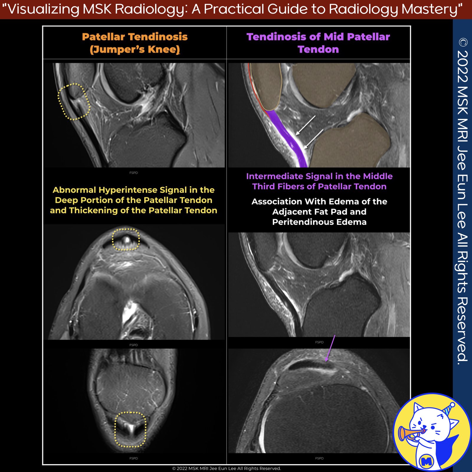
📌 Patellar Tendinosis
✅ Overview
- Patellar tendinosis, also known as jumper's knee, is a common disorder affecting the patellar tendon, particularly in young, active individuals.
- Terminology for overuse-related patellar tendon diseases is inconsistent, with terms such as tendinitis, tendinosis, and partial insertional tear used interchangeably to describe abnormalities near the patellar attachment.
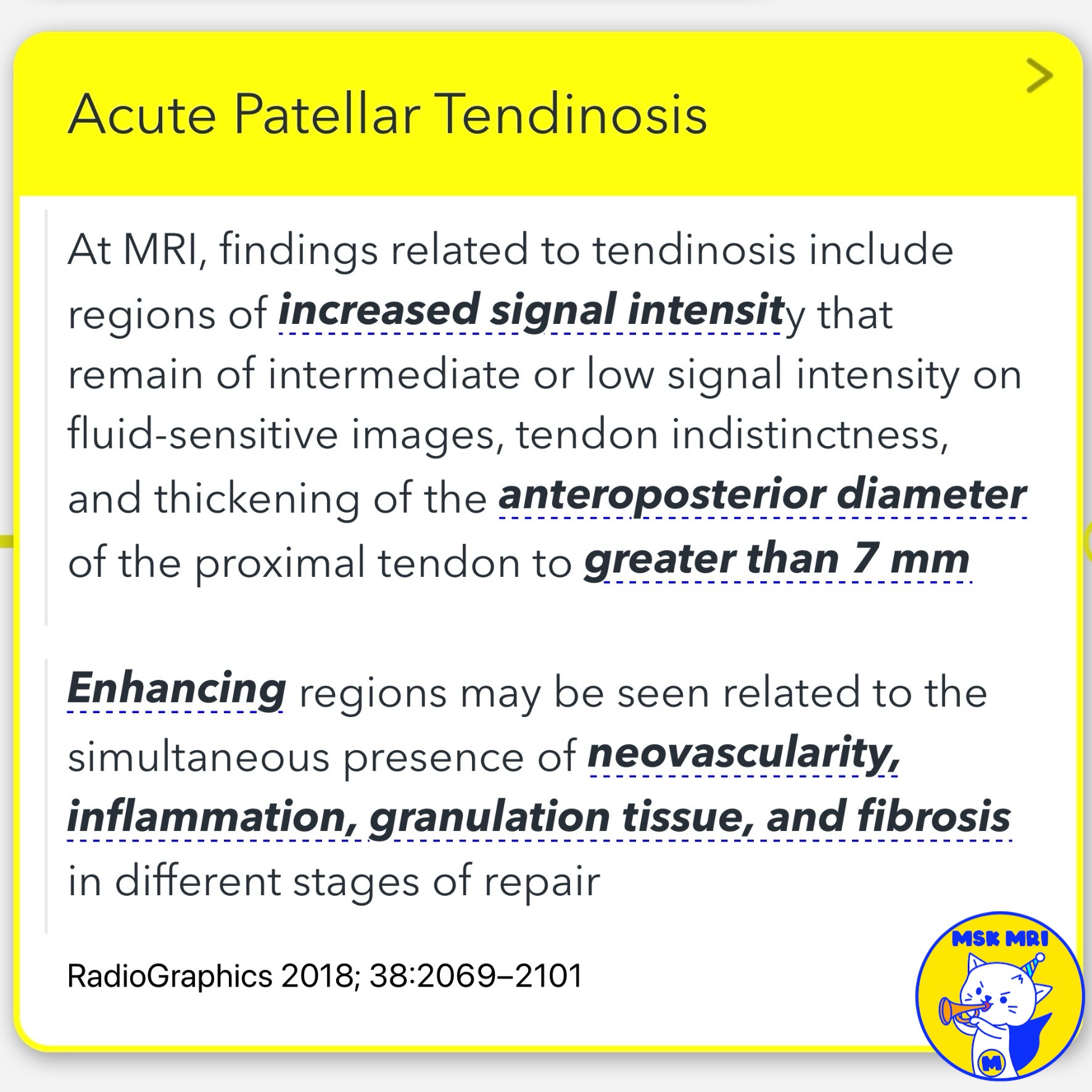
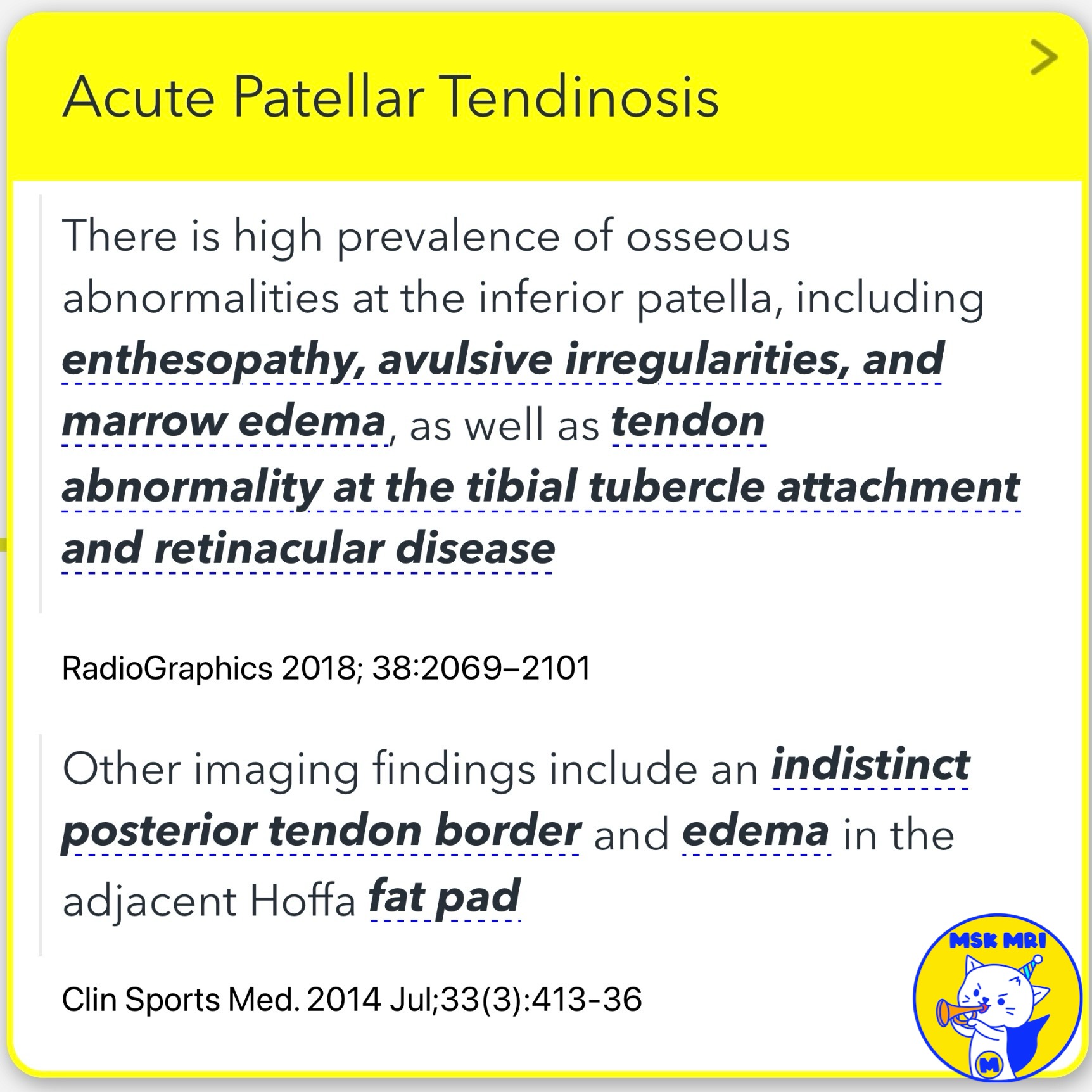
1️⃣ Acute Patellar Tendinosis
In acute cases, symptoms last for less than two weeks. MRI findings include:
- Increased signal intensity on fluid-sensitive images
- Tendon indistinctness and thickening
- Enhancing regions indicating neovascularity, inflammation, granulation tissue, and fibrosis
- Osseous abnormalities at the inferior patella, including enthesopathy and marrow edema
- Indistinct posterior tendon border and edema in Hoffa's fat pad
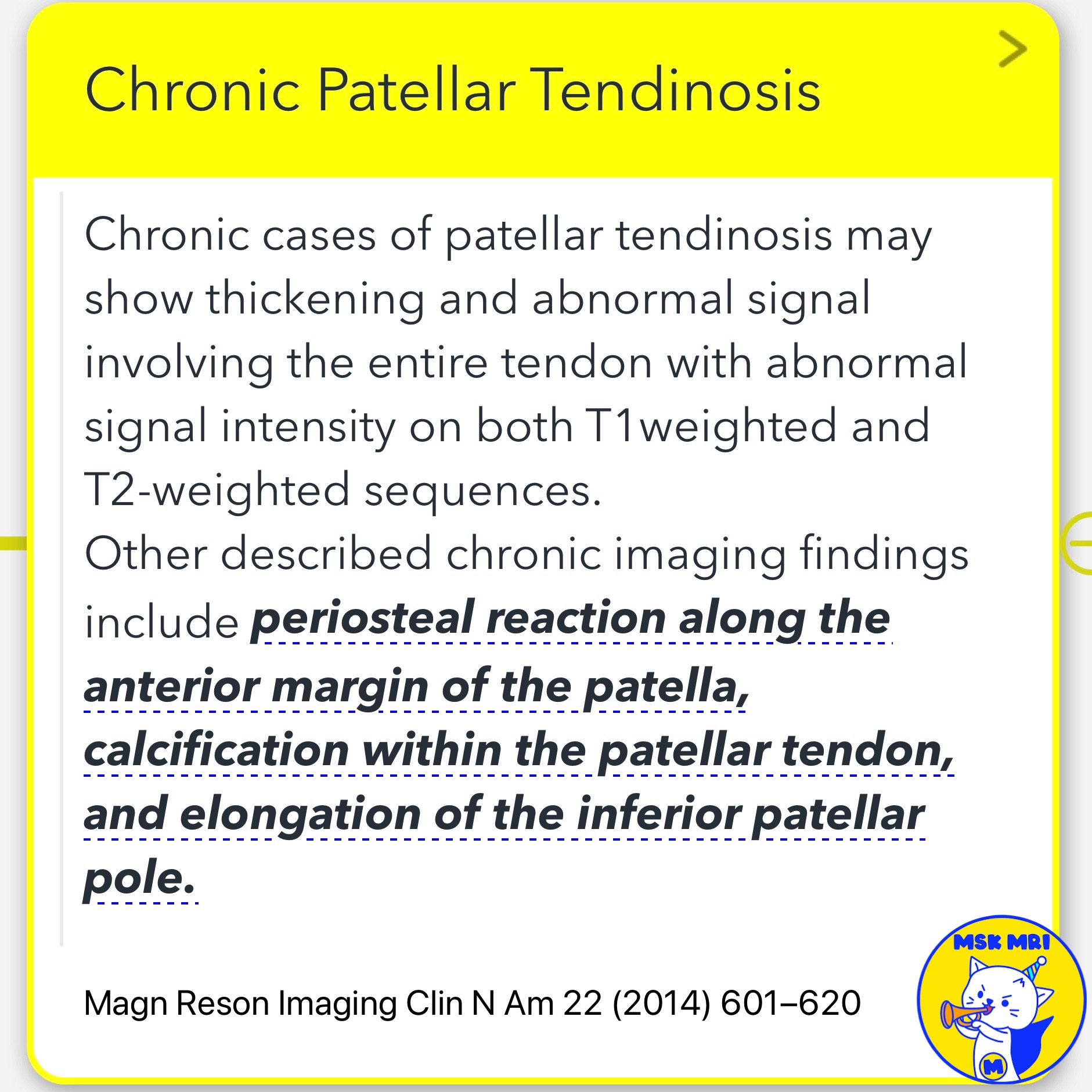
2️⃣ Chronic Patellar Tendinosis
Chronic cases, with symptoms lasting more than six weeks, show:
- Thickening and abnormal signal in the entire tendon on T1 and T2-weighted sequences
- Periosteal reaction along the patella
- Calcification within the tendon
- Elongation of the inferior patellar pole
- Diffuse tendon thickening or enlargement in severe cases
📌Patellar Tendon Tear
- Though infrequent, patellar tendon tears typically occur in adult men under 40 years old due to high-velocity eccentric contraction of the quadriceps.
- Predisposing factors include chronic microtrauma, previous tendon grafts, prolonged steroid use, and systemic diseases.
- Complete tears are less common than partial tears and usually occur at the patellar attachment, showing extensive edema in surrounding soft tissues.
References
- Magn Reson Imaging Clin N Am 22 (2014) 601–620
- RadioGraphics 2018; 38:2069–2101
- Clin Sports Med. 2014 Jul;33(3):413-36
- Radiographics. 2009 May-Jun;29(3):877-86
- Eur J Radiol. 2007 Apr;62(1):27-43
- Stoller's Orthopaedics and Sports Medicine, David W. Stoller MD FACR, LWW, 2016
"Visualizing MSK Radiology: A Practical Guide to Radiology Mastery"
© 2022 MSK MRI Jee Eun Lee All Rights Reserved.
No unauthorized reproduction, redistribution, or use for AI training.
#jumperknee, #patellartendinosis, #patellartendinitis, #kneepain, #sportsinjuries, #MRI, #tendonhealth, #orthopedics, #radiology, #youthathletes


'✅ Knee MRI Mastery > Chap 4BCD. Anterior knee' 카테고리의 다른 글
| (Fig 4-B.16) Osgood Schlatter Disease: Cartilaginous Stage (1) | 2024.06.15 |
|---|---|
| (Fig 4-B.15) Patellar Tendon Partial Tear (0) | 2024.06.12 |
| (Fig 4-B.13) Traumatic Separation of Prepatellar Quadriceps Continuation (0) | 2024.06.12 |
| (Fig 4-B.12) Quadriceps Tendinosis (0) | 2024.06.12 |
| (Fig 4-B.11) Quadriceps Tendon Partial Tear (0) | 2024.06.12 |