Click the link to purchase on Amazon 🎉📚
==============================================
🎥 Check Out All Videos at Once! 📺
👉 Visit Visualizing MSK Blog to explore a wide range of videos! 🩻
https://visualizingmsk.blogspot.com/?view=magazine
📚 You can also find them on MSK MRI Blog and Naver Blog! 📖
https://www.instagram.com/msk_mri/
Click now to stay updated with the latest content! 🔍✨
==============================================
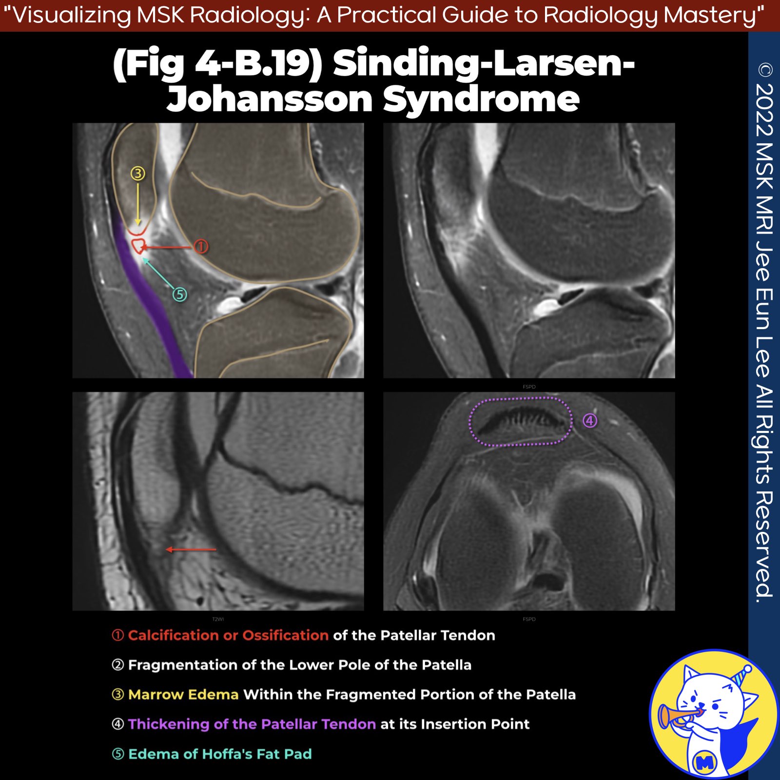
📌Sinding-Larsen-Johansson Syndrome
- Sinding-Larsen-Johansson syndrome, also known as jumper's knee, is caused by repetitive microtrauma to the patellar tendon at its attachment on the inferior pole of the patella.
✅ Etiology
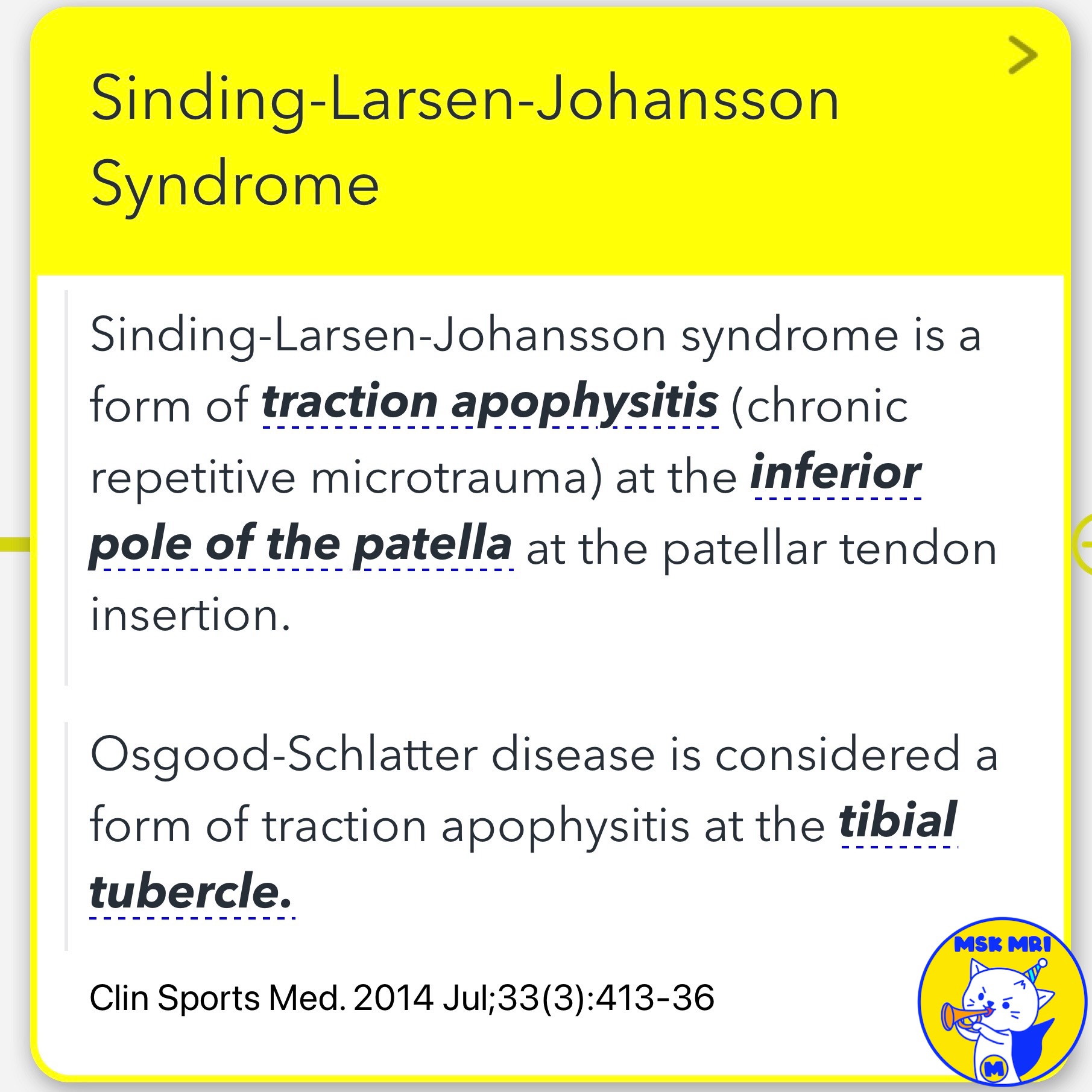
- This syndrome is a type of traction apophysitis that occurs at the inferior pole of the patella.
- In contrast, Osgood-Schlatter disease is traction apophysitis at the tibial tubercle.
✅ Imaging Findings
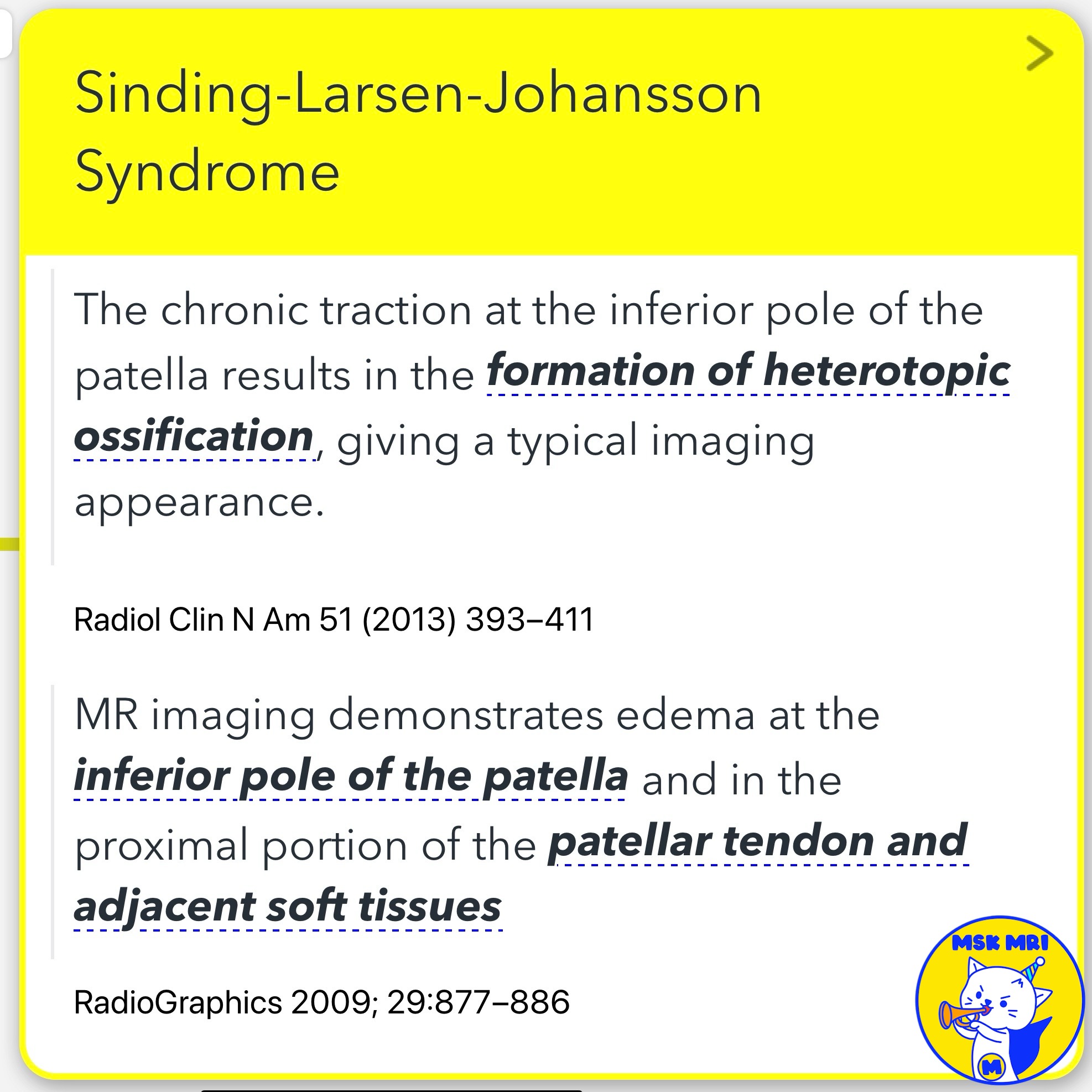
- Chronic traction can lead to heterotopic ossification, which is identifiable through imaging.
- MRI typically shows edema at the inferior pole of the patella and in the proximal portion of the patellar tendon and surrounding soft tissues.
References
- RadioGraphics 2009; 29:877–886
- Clin Sports Med. 2014 Jul;33(3):413-36
- Radiol Clin N Am 51 (2013) 393–411
"Visualizing MSK Radiology: A Practical Guide to Radiology Mastery"
© 2022 MSK MRI Jee Eun Lee All Rights Reserved.
No unauthorized reproduction, redistribution, or use for AI training.
#SindingLarsenJohanssonSyndrome, #JumpersKnee, #TractionApophysitis, #PatellarTendon, #Microtrauma, #HeterotopicOssification, #MRI, #SportsInjuries, #Orthopedics, #Radiology


'✅ Knee MRI Mastery > Chap 4BCD. Anterior knee' 카테고리의 다른 글
| (Fig 4-B.21) Patellar Sleeve Fracture and Repair (1) | 2024.06.15 |
|---|---|
| (Fig 4-B.20) Patellar Sleeve Fracture at the Superior Pole (0) | 2024.06.15 |
| (Fig 4-B.18) Osgood Schlatter Disease: Chronic Active Stage (1) | 2024.06.15 |
| (Fig 4-B.17) Subchondral Fracture vs. Osgood Schlatter Disease (0) | 2024.06.15 |
| (Fig 4-B.16) Osgood Schlatter Disease: Cartilaginous Stage (1) | 2024.06.15 |