Click the link to purchase on Amazon 🎉📚
==============================================
🎥 Check Out All Videos at Once! 📺
👉 Visit Visualizing MSK Blog to explore a wide range of videos! 🩻
https://visualizingmsk.blogspot.com/?view=magazine
📚 You can also find them on MSK MRI Blog and Naver Blog! 📖
https://www.instagram.com/msk_mri/
Click now to stay updated with the latest content! 🔍✨
==============================================
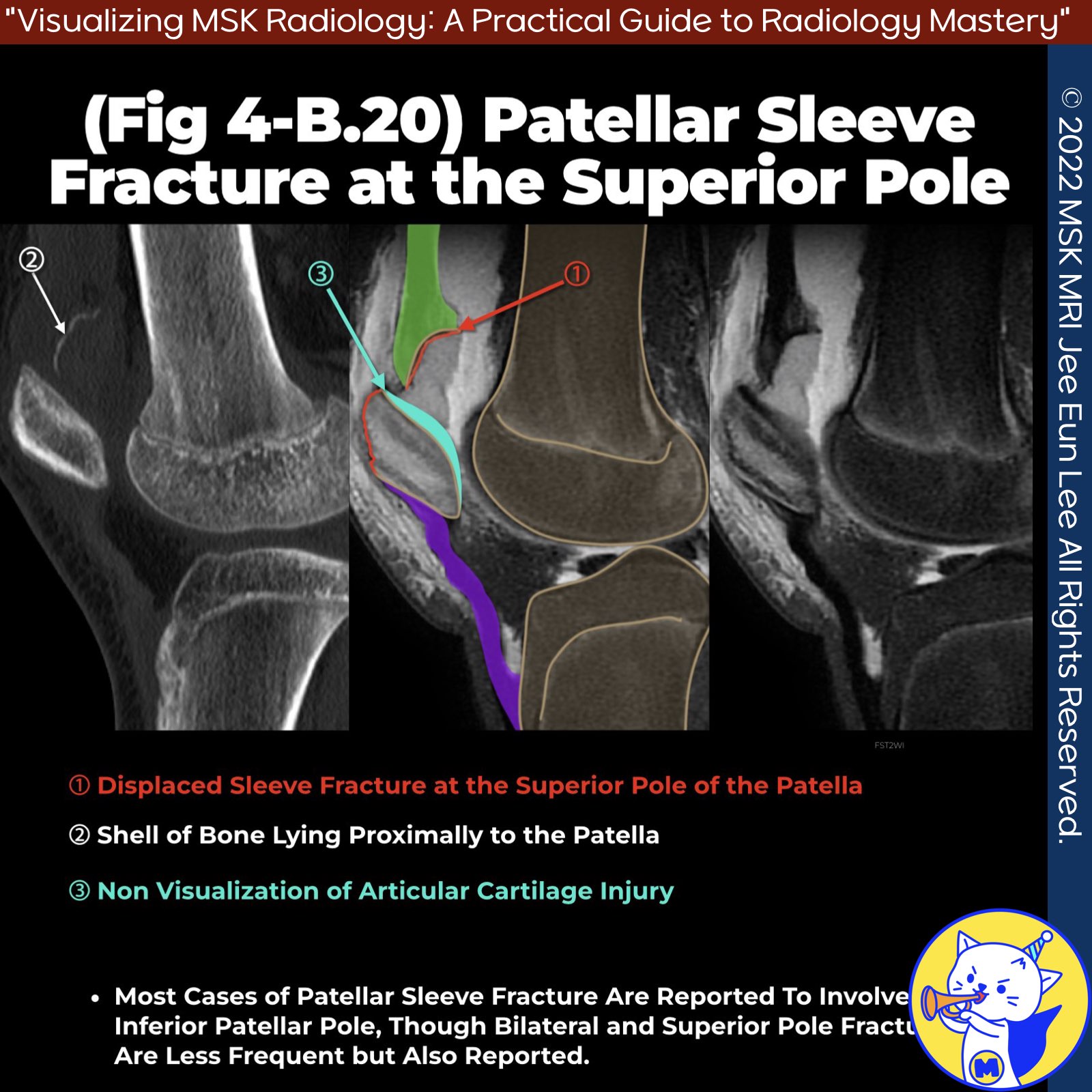
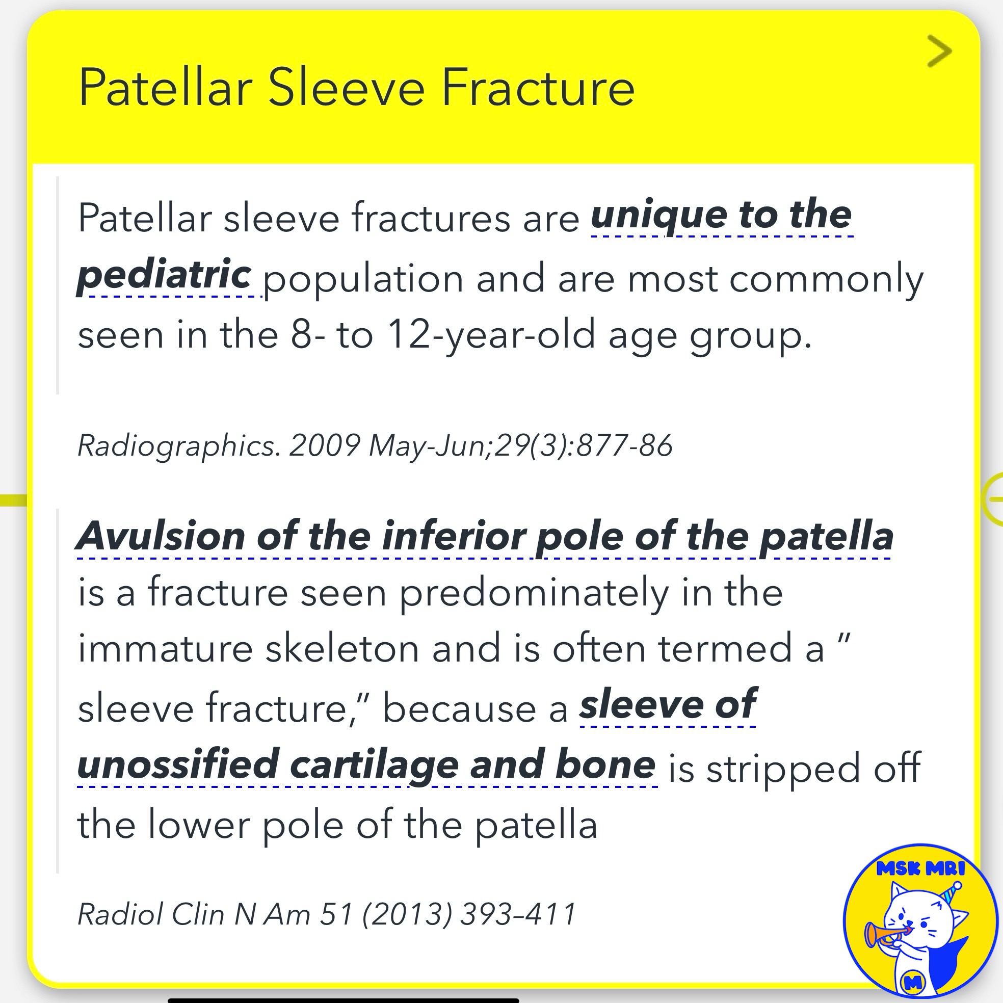
📌 Patellar Sleeve Fracture
- Peak incidence at 12.7 years, ranging from 8 to 16 years.
- Most cases involve the inferior patellar pole, but bilateral and superior pole fractures are also reported.
- These fractures may occur in the entire circumference of the patella and are described as superior, inferior, medial, and lateral avulsions.
✅ Pathophysiology
- This occurs due to the patella's hypermobility and the high cartilage-bone ratio at the transformation zone, which makes it vulnerable to acute and chronic eccentric loads and shear forces.
- The cartilage blends directly with the tendon collagen rather than through distinct Sharpey fibers found in adults.
✅Plain Radiographs
- Demonstrate a small bone fragment inferior to the lower pole of the patella, patella alta, and joint effusion.
- Avulsed ossified fragments are associated with prepatellar swelling and may extend posteriorly to involve the articular surface.
- Radiographs often underestimate the extent of injury at the non-mineralized cartilage and perichondrium.
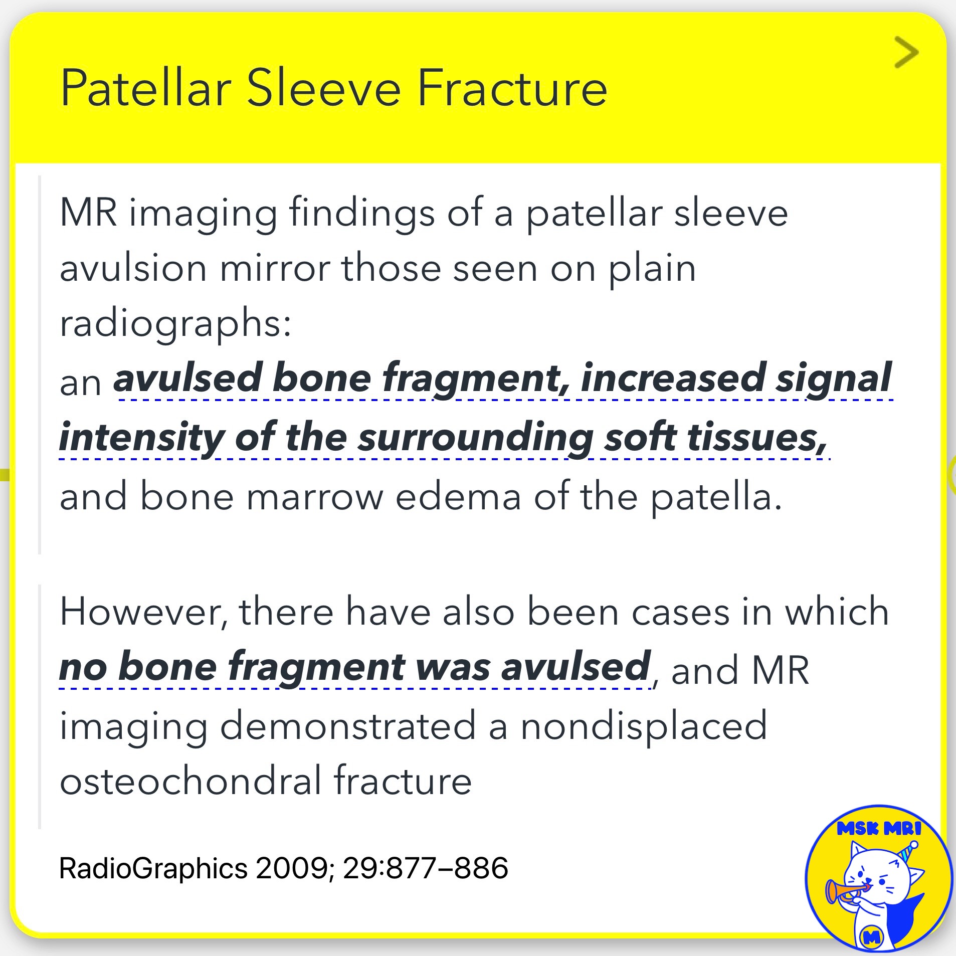
✅ MRI Findings
- Shows an avulsed bone fragment, increased signal intensity of the surrounding soft tissues, and bone marrow edema of the patella.
- MRI can demonstrate a nondisplaced osteochondral fracture if no bone fragment is avulsed.
- A joint effusion may be present if the fracture extends into the joint.
✅ Differential Diagnosis
- Shares imaging features with Sinding-Larsen-Johansson syndrome but has a different clinical manifestation, with an acute onset after a single episode of trauma.
References
- Radiographics. 2009 May-Jun;29(3):877-86
- Radiol Clin N Am 51 (2013) 393–411
- BMC Musculoskelet Disord. 2020 Apr 23;21(1):267
- Knee. 2005 Jan;12(1):3-7
- RadioGraphics 2018; 38:2069–2101
"Visualizing MSK Radiology: A Practical Guide to Radiology Mastery"
© 2022 MSK MRI Jee Eun Lee All Rights Reserved.
No unauthorized reproduction, redistribution, or use for AI training.
#PatellarFracture, #PediatricOrthopedics, #MusculoskeletalRadiology, #AvulsionFracture, #Patella, #Radiology, #MRI, #OrthopedicInjuries, #ChildrensHealth, #TraumaImaging


'✅ Knee MRI Mastery > Chap 4BCD. Anterior knee' 카테고리의 다른 글
| (Fig 4-B.22) Modified Ogden Classification (0) | 2024.06.15 |
|---|---|
| (Fig 4-B.21) Patellar Sleeve Fracture and Repair (1) | 2024.06.15 |
| (Fig 4-B.19) Sinding-Larsen-Johansson Syndrome (1) | 2024.06.15 |
| (Fig 4-B.18) Osgood Schlatter Disease: Chronic Active Stage (1) | 2024.06.15 |
| (Fig 4-B.17) Subchondral Fracture vs. Osgood Schlatter Disease (0) | 2024.06.15 |