Click the link to purchase on Amazon 🎉📚
==============================================
🎥 Check Out All Videos at Once! 📺
👉 Visit Visualizing MSK Blog to explore a wide range of videos! 🩻
https://visualizingmsk.blogspot.com/?view=magazine
📚 You can also find them on MSK MRI Blog and Naver Blog! 📖
https://www.instagram.com/msk_mri/
Click now to stay updated with the latest content! 🔍✨
==============================================
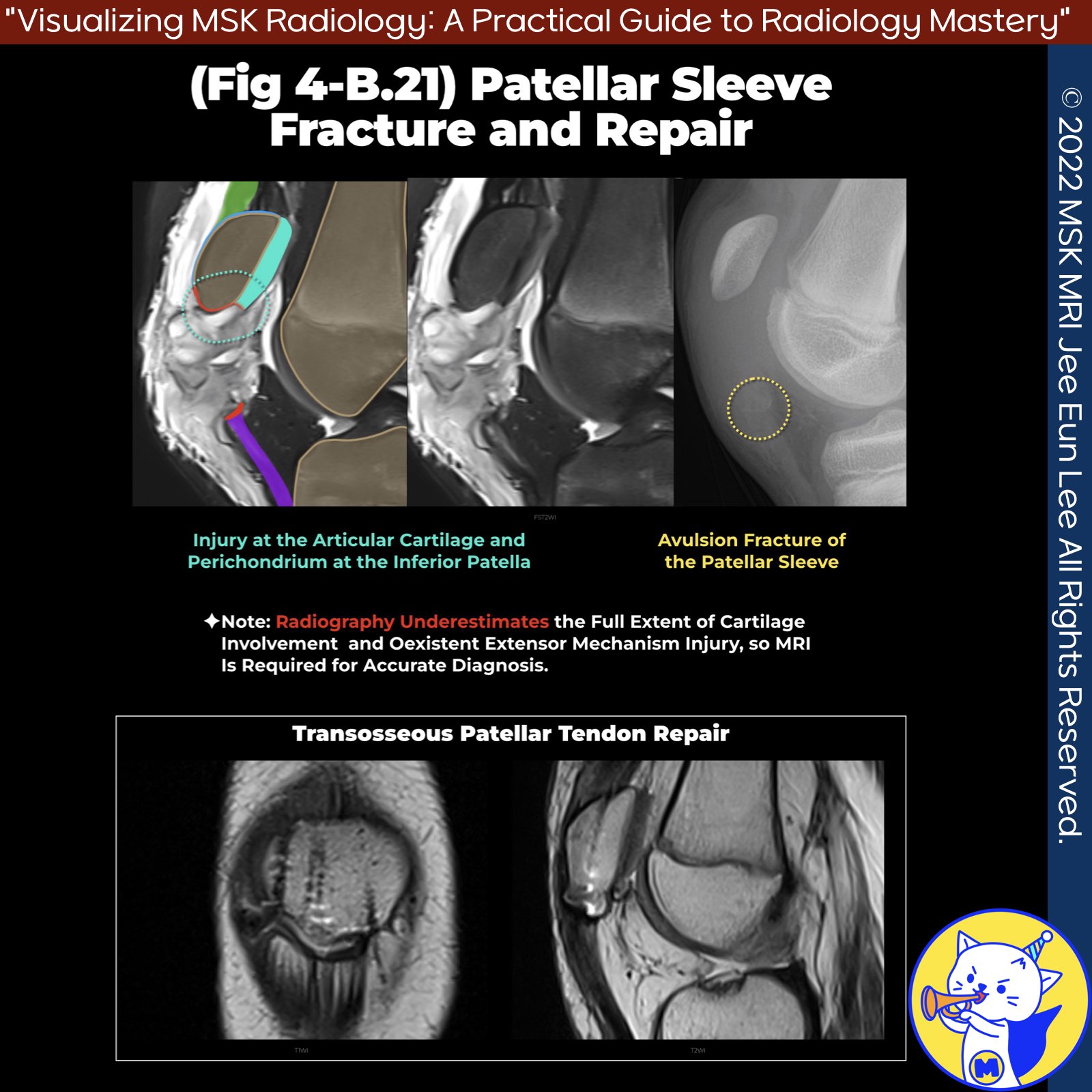
📌Patellar Sleeve Avulsion Fracture: A Case Study
✅ Radiographic Findings:
- Patella alta
- Joint effusion
- Visible Hoffa's fat pad and patellar tendon
- Linear high-density structure near tendon's proximal attachment
✅ MRI Findings:
- Clear injury to articular cartilage and perichondrium of the inferior patella
- Hematoma filling the injured space
✅ Anatomical Notes:
- The posterior patella mostly covered by hyaline cartilage
- Inferior-most part lacks cartilage
- Acute traumatic chondral injury evident
✅ Key Point:
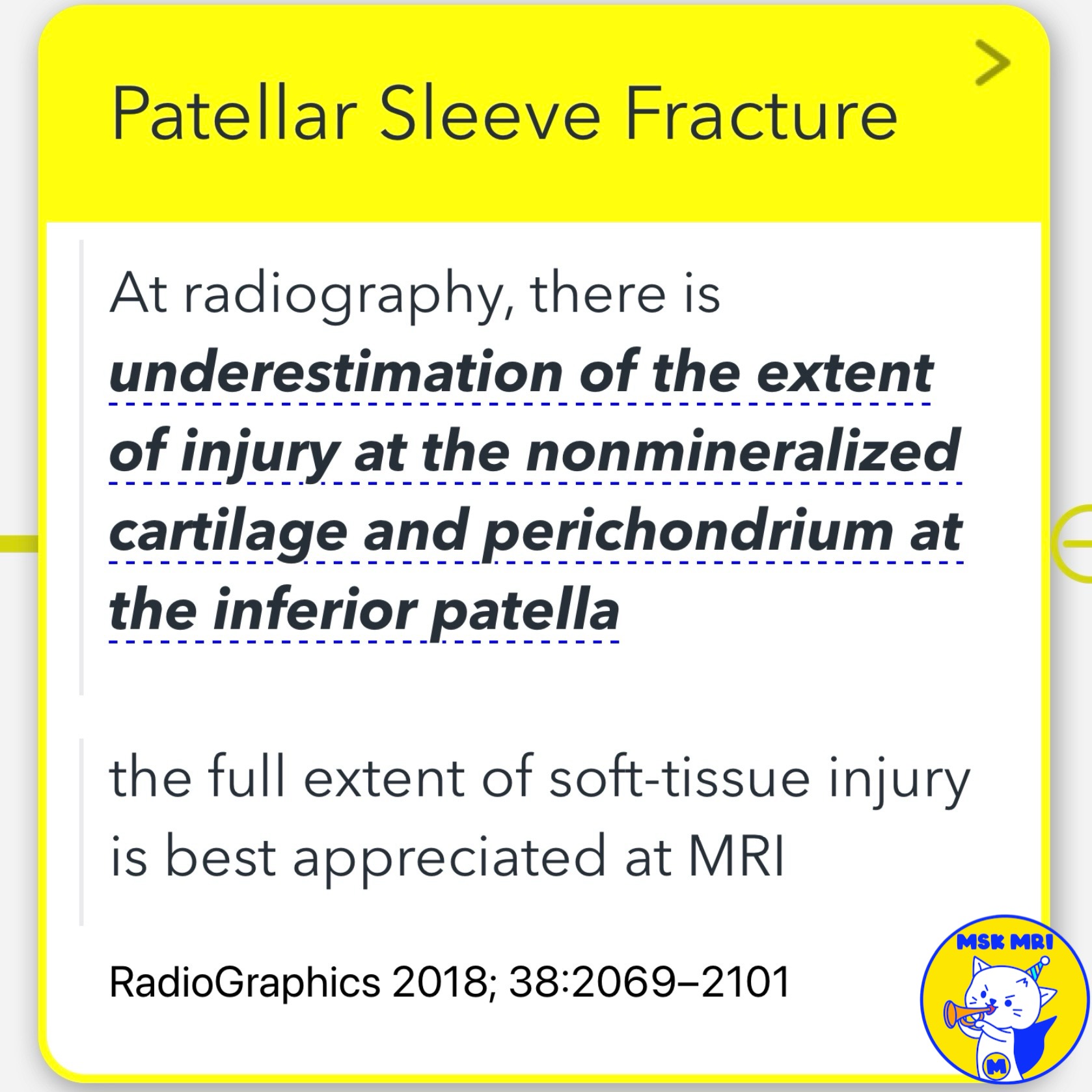
- Radiographs often underestimate injury extent in nonmineralized cartilage and perichondrium
✅ Management:
- Operative intervention with anatomic transosseous suture repair
- Excellent patient outcomes
"Visualizing MSK Radiology: A Practical Guide to Radiology Mastery"
© 2022 MSK MRI Jee Eun Lee All Rights Reserved.
No unauthorized reproduction, redistribution, or use for AI training.
#PatellarSleeveFracture, #OrthopedicImaging, #MRIFindings, #KneeInjury, #SportsTrauma, #PediatricOrthopedics, #RadiologyEducation, #SurgicalManagement, #AnatomicRepair, #ClinicalOutcomes


'✅ Knee MRI Mastery > Chap 4BCD. Anterior knee' 카테고리의 다른 글
| (Fig 4-B.23) Ogden Type IA Tibial Tuberosity Fracture (0) | 2024.06.15 |
|---|---|
| (Fig 4-B.22) Modified Ogden Classification (0) | 2024.06.15 |
| (Fig 4-B.20) Patellar Sleeve Fracture at the Superior Pole (0) | 2024.06.15 |
| (Fig 4-B.19) Sinding-Larsen-Johansson Syndrome (1) | 2024.06.15 |
| (Fig 4-B.18) Osgood Schlatter Disease: Chronic Active Stage (1) | 2024.06.15 |