==============================================
🎥 Check Out All Videos at Once! 📺
👉 Visit Visualizing MSK Blog to explore a wide range of videos! 🩻
https://visualizingmsk.blogspot.com/?view=magazine
📚 You can also find them on MSK MRI Blog and Naver Blog! 📖
https://www.instagram.com/msk_mri/
Click now to stay updated with the latest content! 🔍✨
==============================================
📌Ogden Classification System for Tibial Tubercle Fractures
The Ogden classification system categorizes tibial tubercle fractures into five types, each with subtypes that describe the extent and nature of the fracture.
1️⃣ Type I Fractures
Type I fractures involve only the distal portion of the tubercle.
- Type IA: Minimally displaced or non-displaced fracture.
- Type IB: Anterior and proximal displacement or comminution.
- Type IC: Proposed subtype for associated patellar tendon avulsions.
References:
- RadioGraphics 2009; 29:877–886
- Open Journal of Medical Imaging, 2013, 3, 90-96
- EFORT Open Rev. 2020 May 5;5(5):260-267
2️⃣ Type II Fractures
Type II fractures involve the entire ossification center of the tubercle.
- Type IIA: Separation of the tubercle from the proximal tibia, minimally or non-displaced.
- Type IIB: Anterior displacement and/or comminution.
References:
- RadioGraphics 2009; 29:877–886
- Open Journal of Medical Imaging, 2013, 3, 90-96
- EFORT Open Rev. 2020 May 5;5(5):260-267
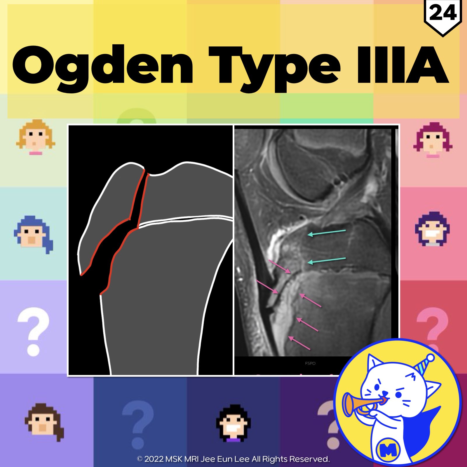
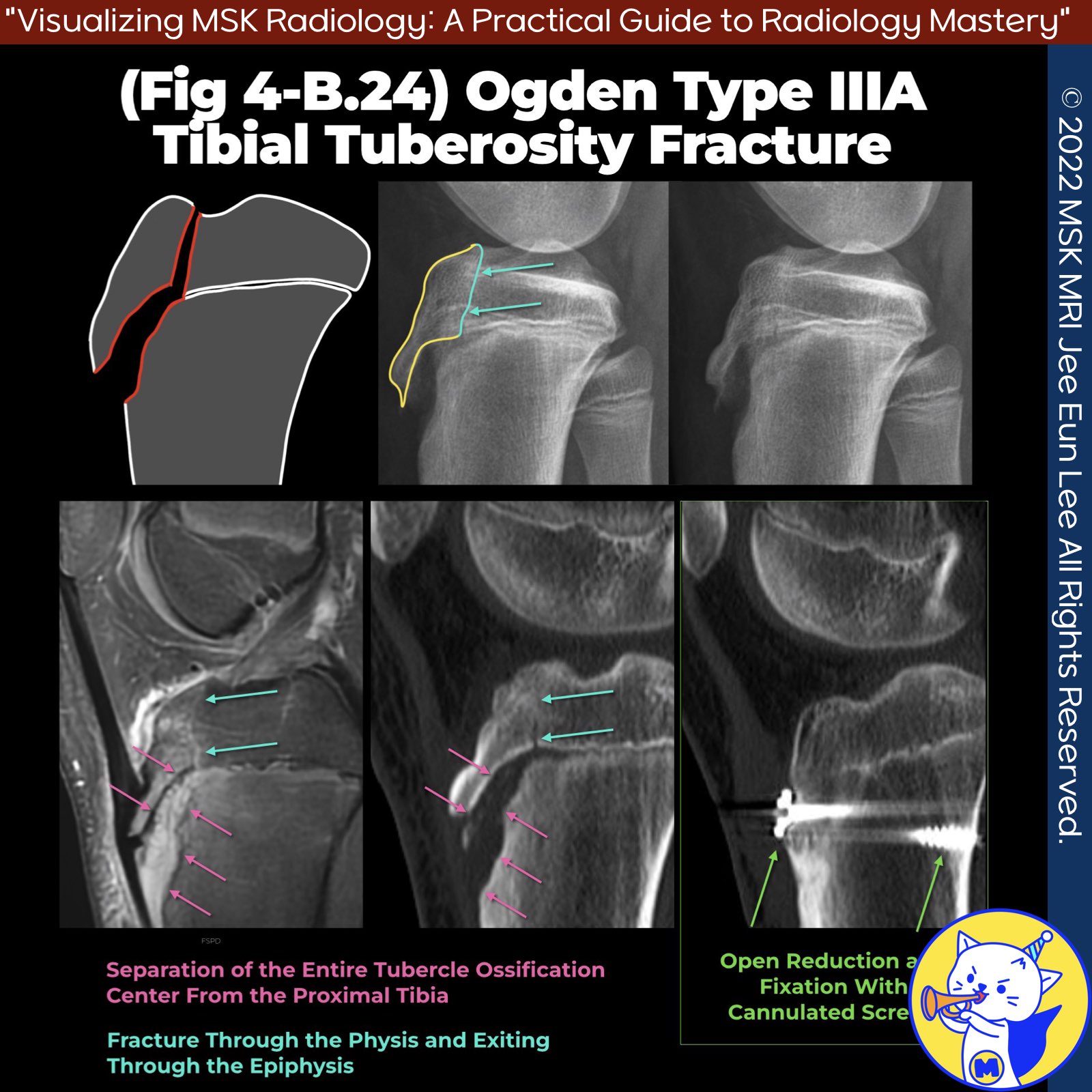
3️⃣ Type III Fractures
Type III fractures extend through the proximal tibial epiphysis into the joint space.
- Type IIIA: Non-displaced fracture.
- Type IIIB: Comminuted fracture.
References:
- RadioGraphics 2009; 29:877–886
- Open Journal of Medical Imaging, 2013, 3, 90-96
- Radiol Clin N Am 51 (2013) 393–411
- EFORT Open Rev. 2020 May 5;5(5):260-267
4️⃣Type IV Fractures
Type IV fractures present with fracture extension from the tibial tuberosity through the proximal tibial physis into the posterior tibial metaphyseal cortex.
References:
- Open Journal of Medical Imaging, 2013, 3, 90-96
- EFORT Open Rev. 2020 May 5;5(5):260-267
5️⃣ Type V Fractures
Type V fractures consist of a combined type III and type IV avulsion fracture, resulting in an inverted “Y” configuration.
References:
- Open Journal of Medical Imaging, 2013, 3, 90-96
- EFORT Open Rev. 2020 May 5;5(5):260-267
Treatment Guidelines
- Type I Fractures: Usually treated with knee immobilization in complete extension.
- Type II and III Fractures: Typically require osseous fixation with pins or screws.
Reference:
- RadioGraphics 2009; 29:877–886
Further Reading
- Journal of Children’s Orthopaedics, Vol. 2, No. 6, 2008, pp. 469-474
- Clinical Orthopaedics and Related Research, No. 194, 1980, pp. 181-184
- Orthopedic Clinics of North America, Vol. 34, No. 3, 2003, pp. 397-403
- Journal of Bone and Joint Surgery (American Volume), Vol. 72, No. 9, 1990, pp. 1411-1413
#TibialFractures, #OgdenClassification, #TypeIFractures, #TypeIIFractures, #TypeIIIFractures, #TypeIVFractures, #TypeVFractures, #Orthopedics, #Radiology, #SportsMedicine

'✅ Knee MRI Mastery > Chap 4BCD. Anterior knee' 카테고리의 다른 글
| (Fig 4-B.26) Pre-Patellar Friction Syndrome (0) | 2024.06.16 |
|---|---|
| (Fig 4-B.25) Ogden Type IV Tibial Tuberosity Fracture (0) | 2024.06.16 |
| (Fig 4-B.23) Ogden Type IA Tibial Tuberosity Fracture (0) | 2024.06.15 |
| (Fig 4-B.22) Modified Ogden Classification (0) | 2024.06.15 |
| (Fig 4-B.21) Patellar Sleeve Fracture and Repair (1) | 2024.06.15 |


