Click the link to purchase on Amazon 🎉📚
==============================================
🎥 Check Out All Videos at Once! 📺
👉 Visit Visualizing MSK Blog to explore a wide range of videos! 🩻
https://visualizingmsk.blogspot.com/?view=magazine
📚 You can also find them on MSK MRI Blog and Naver Blog! 📖
https://www.instagram.com/msk_mri/
Click now to stay updated with the latest content! 🔍✨
==============================================
📌 Pre-Patellar Friction Syndrome
✅ Anatomy:
- Superficial Layer: Transverse orientation to the patella.
- Intermediate Layer: Oblique orientation, arising from the expansion of the vastus medialis and lateralis with some contribution from the superficial fibers of rectus femoris.
- Deep Layer: The Prepatellar Quadriceps Continuation, Longitudinal orientation, formed by the deep fibers of rectus femoris, adherent to the underlying bone, and continuing inferiorly to become part of the patellar tendon.
✅ Clinical Features:
- Anterior knee pain during physical exertion.
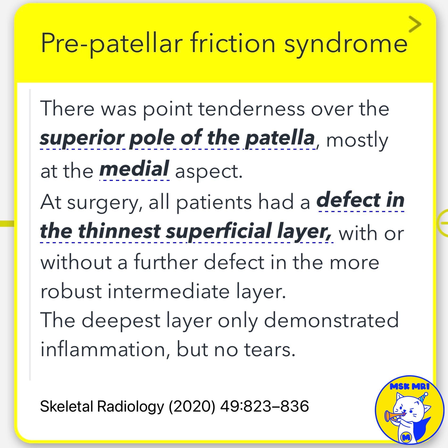
- Point tenderness over the superior pole of the patella, mostly at the medial aspect.
- Caused by repetitive motion leading to the superficial and intermediate layers of the pre-patellar soft tissues rubbing against the fixed deep layer.
✅ Diagnosis:
- Ultrasound: Demonstrates fascial defects.
- MRI: Expected to show oedematous swelling of the pre-patellar soft tissues.
Defects in the thinnest superficial layer observed during surgery, with possible defects in the intermediate layer.
Inflammation in the deepest layer without tears. - ★ Important to differentiate from pre-patellar bursitis on MRI.
Skeletal Radiology (2020) 49:823–836
"Visualizing MSK Radiology: A Practical Guide to Radiology Mastery"
© 2022 MSK MRI Jee Eun Lee All Rights Reserved.
No unauthorized reproduction, redistribution, or use for AI training.
#PrePatellarFrictionSyndrome, #KneePain, #CyclingInjury, #AnteriorKneePain, #MRI, #Ultrasound, #OrthopedicSurgery, #SportsInjury, #KneeAnatomy, #MedicalResearch
'✅ Knee MRI Mastery > Chap 4BCD. Anterior knee' 카테고리의 다른 글
| (Fig 4-C.02) Patellar Tendon-Lateral Femoral Condyle Friction (0) | 2024.06.17 |
|---|---|
| (Fig 4-C.01) Fat Pads Around the Knee (1) | 2024.06.16 |
| (Fig 4-B.25) Ogden Type IV Tibial Tuberosity Fracture (0) | 2024.06.16 |
| (Fig 4-B.24) Ogden Type IIIA Tibial Tuberosity Fracture (1) | 2024.06.15 |
| (Fig 4-B.23) Ogden Type IA Tibial Tuberosity Fracture (0) | 2024.06.15 |


