==============================================
⬇️✨⬇️🎉⬇️🔥⬇️📚⬇️
Click the link to purchase on Amazon 🎉📚
==============================================
🎥 Check Out All Videos at Once! 📺
👉 Visit Visualizing MSK Blog to explore a wide range of videos! 🩻
https://visualizingmsk.blogspot.com/?view=magazine
📚 You can also find them on MSK MRI Blog and Naver Blog! 📖
https://www.instagram.com/msk_mri/
Click now to stay updated with the latest content! 🔍✨
==============================================
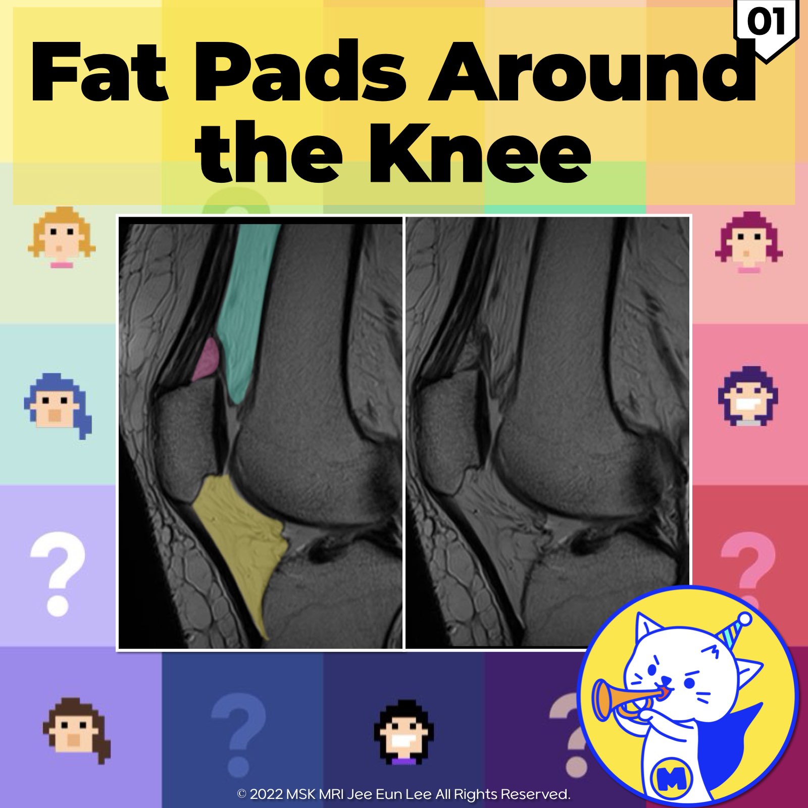
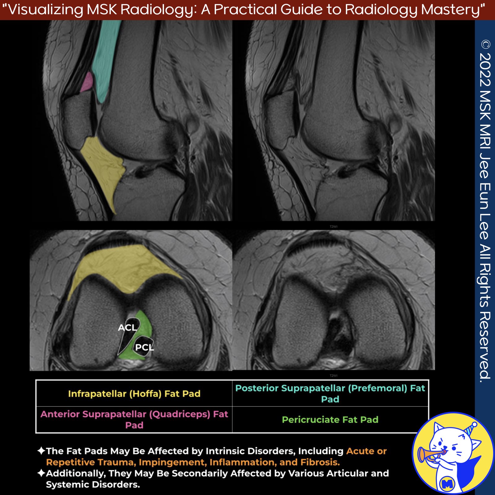
📌 Anatomy of the Fat Pads
- The fat pads of the knee are subject to various intrinsic disorders such as acute or repetitive trauma, impingement, inflammation, and fibrosis.
- These structures may also be secondarily affected by a range of articular and systemic disorders.
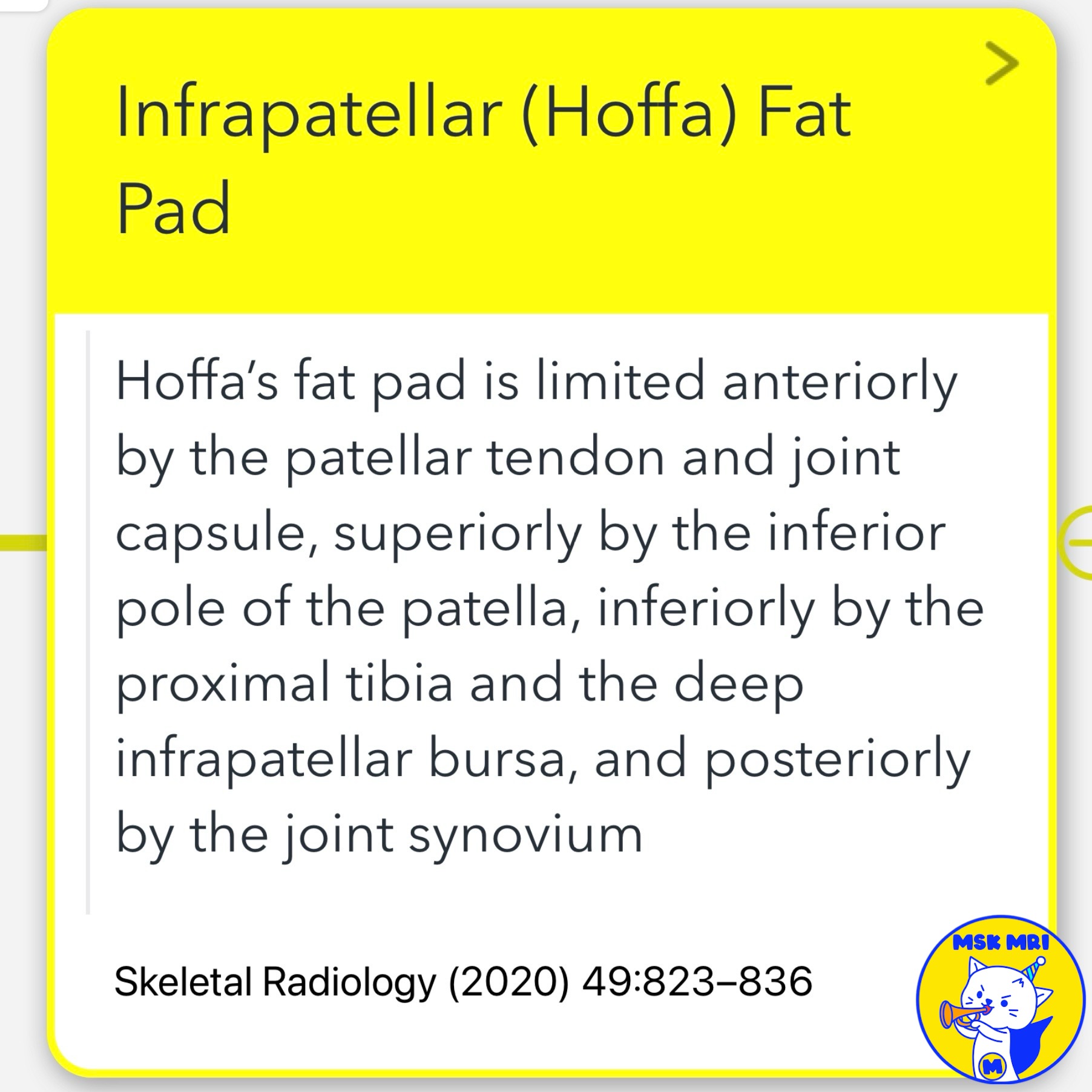
1️⃣. Infrapatellar (Hoffa) Fat Pad
- Hoffa's fat pad is located anteriorly by the patellar tendon and joint capsule, superiorly by the inferior pole of the patella, inferiorly by the proximal tibia and the deep infrapatellar bursa, and posteriorly by the joint synovium.
- It is the most sensitive tissue in the knee, richly innervated by branches of the femoral, common peroneal, and saphenous nerves.
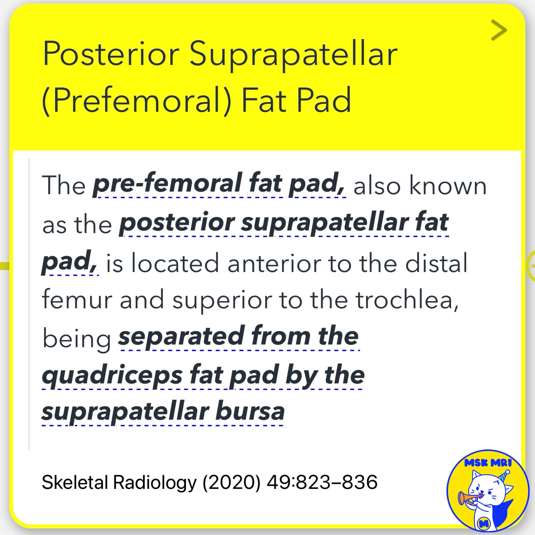
2️⃣. Posterior Suprapatellar (Prefemoral) Fat Pad
- The posterior suprapatellar fat pad, also known as the prefemoral fat pad, is situated anterior to the distal femur and superior to the trochlea.
- It is separated from the quadriceps fat pad by the suprapatellar bursa.
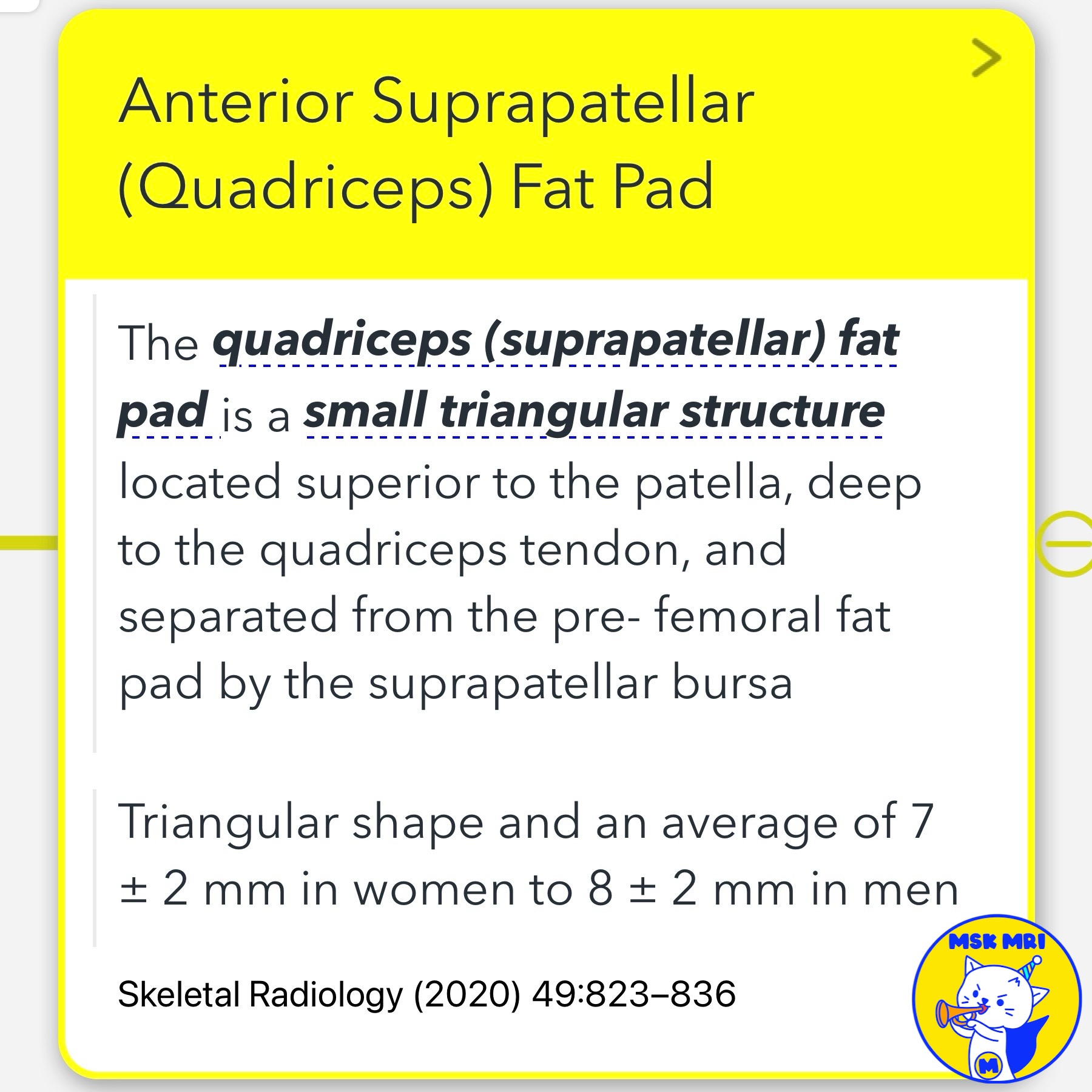
3️⃣. Anterior Suprapatellar (Quadriceps) Fat Pad
- The quadriceps fat pad is a small triangular structure located superior to the patella and deep to the quadriceps tendon.
- It is separated from the prefemoral fat pad by the suprapatellar bursa. This fat pad is believed to promote stress dissipation or act as a mechanosensory organ.
4️⃣. Pericruciate Fat Pad
- The pericruciate fat pad fills the gap between the anterior and posterior cruciate ligaments.
- Similar to the anterior fat pads of the knee, it is intracapsular and extrasynovial.
- It appears as a triangular-shaped structure in the sagittal plane, located above the posterior cruciate ligament and posterior to the fibers of the anterior cruciate ligament.
References
- RadioGraphics 2018; 38:2069–2101
- Skeletal Radiology (2020) 49:823–836
- Magn Reson Imaging Clin N Am 22 (2014) 725–741
"Visualizing MSK Radiology: A Practical Guide to Radiology Mastery"
© 2022 MSK MRI Jee Eun Lee All Rights Reserved.
No unauthorized reproduction, redistribution, or use for AI training.
#KneeAnatomy, #FatPads, #HoffasFatPad, #SuprapatellarFatPad, #PrefemoralFatPad, #QuadricepsFatPad, #PericruciateFatPad, #KneePain, #Radiology, #MSKImaging


'✅ Knee MRI Mastery > Chap 4BCD. Anterior knee' 카테고리의 다른 글
| (Fig 4-C.03) Post-traumatic Lesions of Hoffa’s Fat Pad (2) | 2024.06.17 |
|---|---|
| (Fig 4-C.02) Patellar Tendon-Lateral Femoral Condyle Friction (0) | 2024.06.17 |
| (Fig 4-B.26) Pre-Patellar Friction Syndrome (0) | 2024.06.16 |
| (Fig 4-B.25) Ogden Type IV Tibial Tuberosity Fracture (0) | 2024.06.16 |
| (Fig 4-B.24) Ogden Type IIIA Tibial Tuberosity Fracture (1) | 2024.06.15 |