Click the link to purchase on Amazon 🎉📚
==============================================
🎥 Check Out All Videos at Once! 📺
👉 Visit Visualizing MSK Blog to explore a wide range of videos! 🩻
https://visualizingmsk.blogspot.com/?view=magazine
📚 You can also find them on MSK MRI Blog and Naver Blog! 📖
https://www.instagram.com/msk_mri/
Click now to stay updated with the latest content! 🔍✨
==============================================
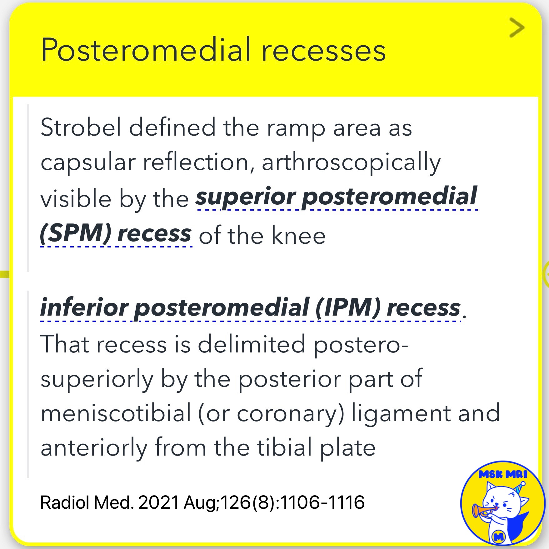
📌 Posteromedial Knee Recesses
✅ Superior Posteromedial Recess
- Borders: Superiorly by the medial femoral condyle, inferiorly by the superior part of the posterior meniscal horn, and posteriorly by the joint capsule.
- Characteristics: When the knee is flexed at 90 degrees during an MRI, this recess widens, while the inferior recess collapses, mimicking the arthroscopic view.
✅ Inferior Posteromedial Recess
- Borders: Posterior part of the meniscotibial ligament and the tibial plateau.
- Characteristics: Typically not accessible with arthroscopy and does not communicate with the superior recess. Prominence can mimic a ramp lesion.
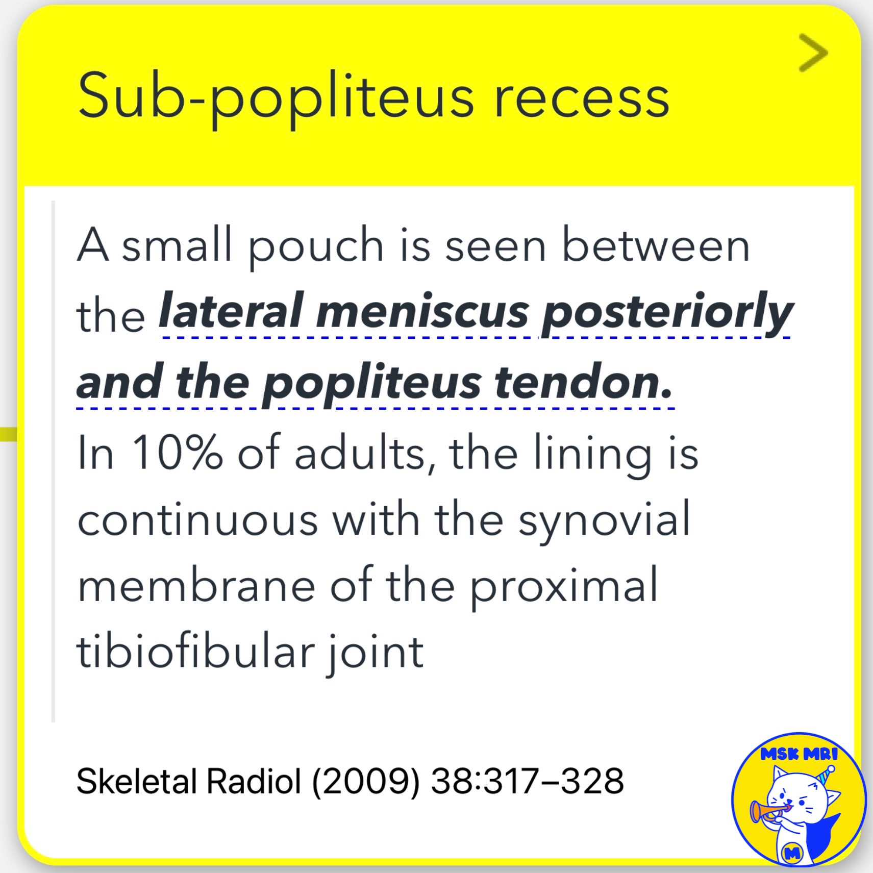
📌 Sub-popliteus (Subpopliteal) Recess
- Located between the popliteus tendon and the posterior horn of the lateral meniscus.
- In about 10% of adults, this lining is continuous with the synovial membrane of the proximal tibiofibular joint.
- Loose bodies can sometimes be found here.
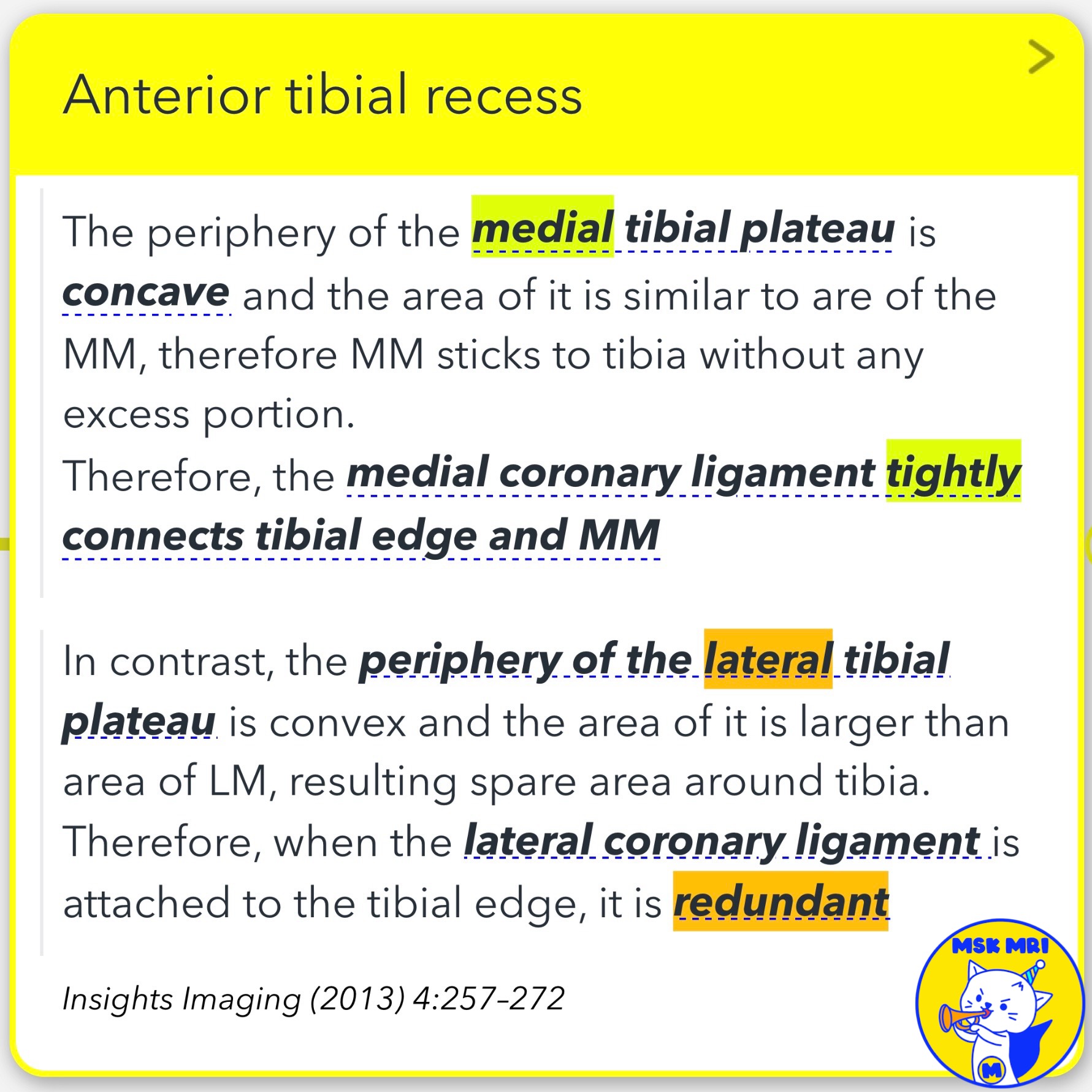
📌 Anterior Tibial Recess
- The anterior tibial recess is a normal capsular recess found just anterior to the proximal tibia.
- It can be observed from both the medial and lateral sides:
✅ Medial Anterior Tibial Recess:
- Smaller due to the tight connection via the medial coronary ligament, matching the concave medial tibial plateau.
✅ Lateral Anterior Tibial Recess:
- Larger due to the redundant lateral coronary ligament, corresponding to the convex and larger lateral tibial plateau.
References
- Insights Imaging (2013) 4:257–272
- J Korean Soc Radiol 2022;83(3):582-596
- Radiol Med. 2021 Aug;126(8):1106-1116
"Visualizing MSK Radiology: A Practical Guide to Radiology Mastery"
© 2022 MSK MRI Jee Eun Lee All Rights Reserved.
No unauthorized reproduction, redistribution, or use for AI training.
#PosteriorMedialCapsule, #MeniscocapsularLigament, #MeniscotibialLigament, #KneeAnatomy, #SagittalMRI, #JointCapsule, #SubpopliteusRecess, #AnteriorTibialRecess, #MedialMeniscus, #KneeMRI

https://www.amazon.com/dp/B0DJGMHMFS

'✅ Knee MRI Mastery > Chap 4BCD. Anterior knee' 카테고리의 다른 글
| (Fig 4-D.16) Inferior Posteromedial Recess vs. Ramp Lesion (0) | 2024.06.26 |
|---|---|
| (Fig 4-D.15) Chondral Loose Bodies in Knee Recesses (0) | 2024.06.26 |
| (Fig 4-D.13) Anatomical Knee Recesses: Part 1 (0) | 2024.06.25 |
| (Fig 4-D.12) Gastrocnemius Ganglia (0) | 2024.06.25 |
| (Fig 4-D.11) Intramuscular Extension of Baker's Cyst (0) | 2024.06.25 |