👉 Click the link below and request access—I’ll approve it for you shortly!
https://www.notion.so/MSKMRI-KNEE-b6cbb1e1bc4741b681ecf6a40159a531?pvs=4
==============================================
✨ Join the channel to enjoy the benefits! 🚀
https://www.youtube.com/channel/UC4bw7o0l2rhxn1GJZGDmT9w/join
==============================================
👉 "Click the link to purchase on Amazon 🎉📚"
[Visualizing MSK Radiology: A Practical Guide to Radiology Mastery]
https://www.amazon.com/dp/B0DJGMHMFS
==============================================
MSK MRI Jee Eun Lee
📚 Visualizing MSK Radiology: A Practical Guide to Radiology Mastery Now! 🌟 Available on Amazon, eBay, and Rain Collectibles! 💻 Ebook coming soon – stay tuned! ⏳ 🔗 https://www.amazon.com/dp/B0DJGMHMFS 🔗 https://www.ebay.com/itm/3875004193
www.youtube.com
Visualizing MSK Radiology: A Practical Guide to Radiology Mastery
www.amazon.com

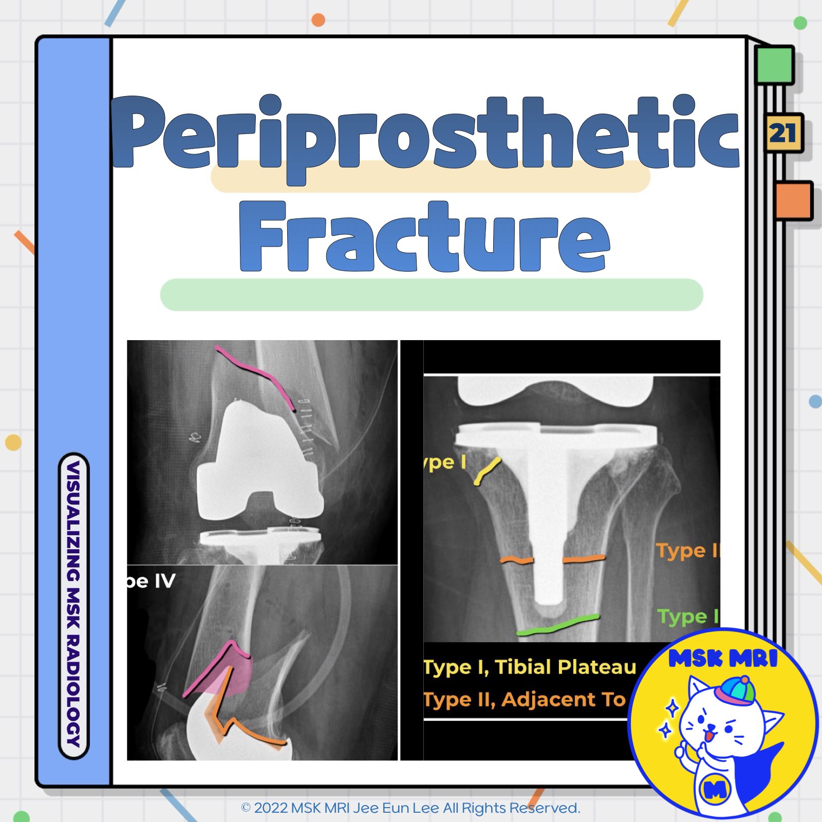
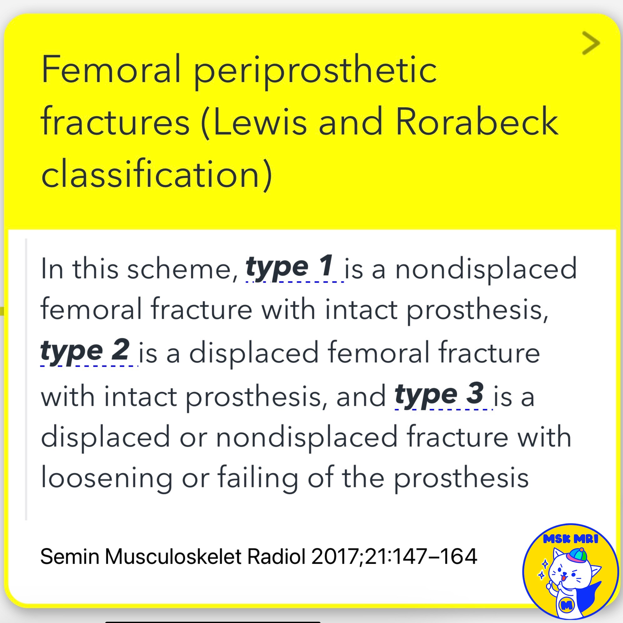
📌 Femoral Periprosthetic Fractures
✅ Lewis and Rorabeck Classification:
- Type 1: Nondisplaced femoral fracture with intact prosthesis
- Type 2: Displaced femoral fracture with intact prosthesis
- Type 3: Displaced or nondisplaced fracture with loosening or failing of the prosthesis
✅ Risk Factors:
- Defects of the anterior femoral cortex from surgical notching
- Cystic lesions of degenerative or rheumatoid origin
- Osteolysis secondary to wear-related debris
✅ Supracondylar Fractures:
- Most frequently encountered fracture near a TKA (Total Knee Arthroplasty)
- Often, a devastating complication
- Classified and managed based on displacement and prosthesis stability
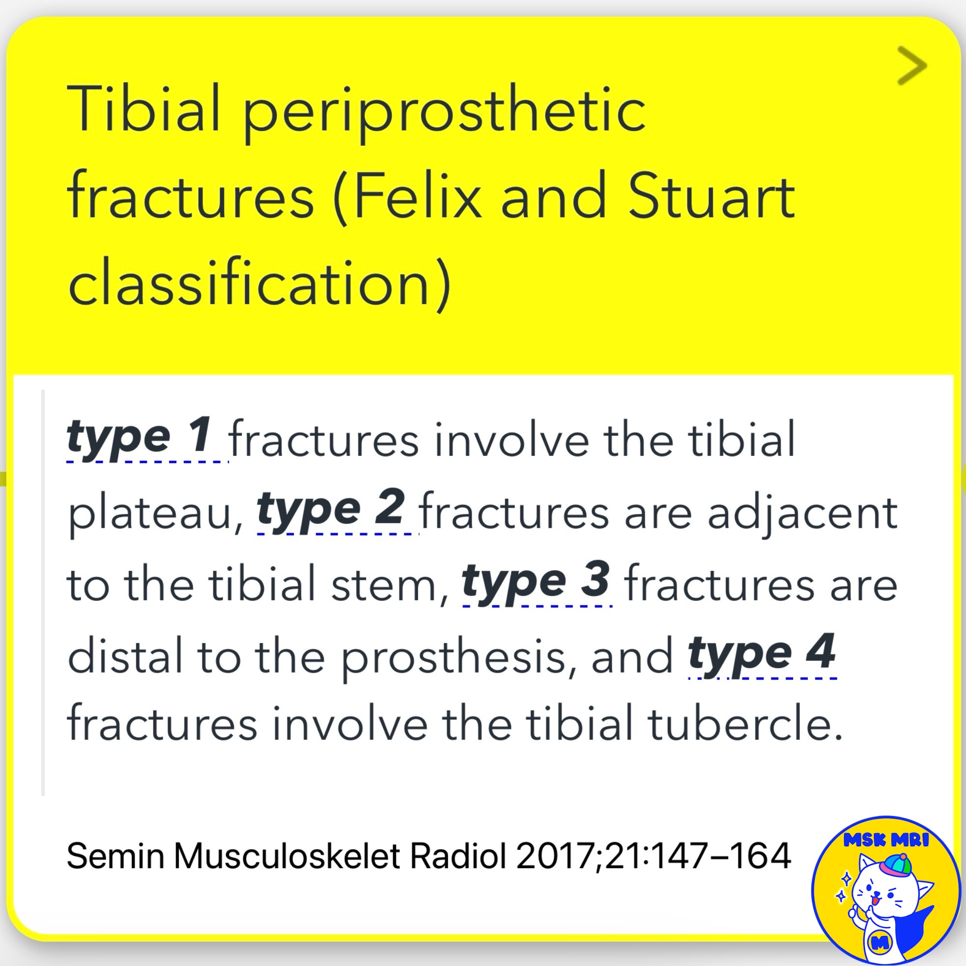
📌 Tibial Periprosthetic Fractures
✅ Felix and Stuart Classification:
- Type 1: Fractures involve the tibial plateau
- Type 2: Fractures are adjacent to the tibial stem
- Type 3: Fractures are distal to the prosthesis
- Type 4: Fractures involve the tibial tubercle
✅ Occurrence and Management:
- Tibial periprosthetic fractures occur infrequently
- Classified and managed based on the anatomic location of the fracture and the stability of the prosthesis
References
- Semin Musculoskelet Radiol 2017;21:147–164
- RadioGraphics 2015; 35:1483–1501
- AJR 2014; 202:W76–W86
"Visualizing MSK Radiology: A Practical Guide to Radiology Mastery"
© 2022 MSK MRI Jee Eun Lee All Rights Reserved.
No unauthorized reproduction, redistribution, or use for AI training.
#PeriprostheticFracture, #FemoralFracture, #TibialFracture, #Orthopedics, #Radiology, #SurgicalComplications, #LewisAndRorabeck, #FelixAndStuart, #TKA, #FractureManagement


'✅ Knee MRI Mastery > Chap 5CD. Cartilage Repair and TKA' 카테고리의 다른 글
| (Fig 5-D.23) Patellar Fracture After TKA (0) | 2024.07.21 |
|---|---|
| (Fig 5-D.22) Varus and Valgus Instability After TKA (0) | 2024.07.21 |
| (Fig 5-D.20) Fracture at Tracking Pin Sites After Robotic-Assisted TKA (0) | 2024.07.21 |
| (Fig 5-D.19) Post-TKA Synovitis: Infection, Particle-Induced, and Nonspecific (0) | 2024.07.21 |
| (Fig 5-D.18) Antibiotic-Loaded Cement CAD-Articulating Spacer (0) | 2024.07.21 |