==============================================
⬇️✨⬇️🎉⬇️🔥⬇️📚⬇️
Click the link to purchase on Amazon 🎉📚
==============================================
🎥 Check Out All Videos at Once! 📺
👉 Visit Visualizing MSK Blog to explore a wide range of videos! 🩻
https://visualizingmsk.blogspot.com/?view=magazine
📚 You can also find them on MSK MRI Blog and Naver Blog! 📖
https://www.instagram.com/msk_mri/
Click now to stay updated with the latest content! 🔍✨
==============================================
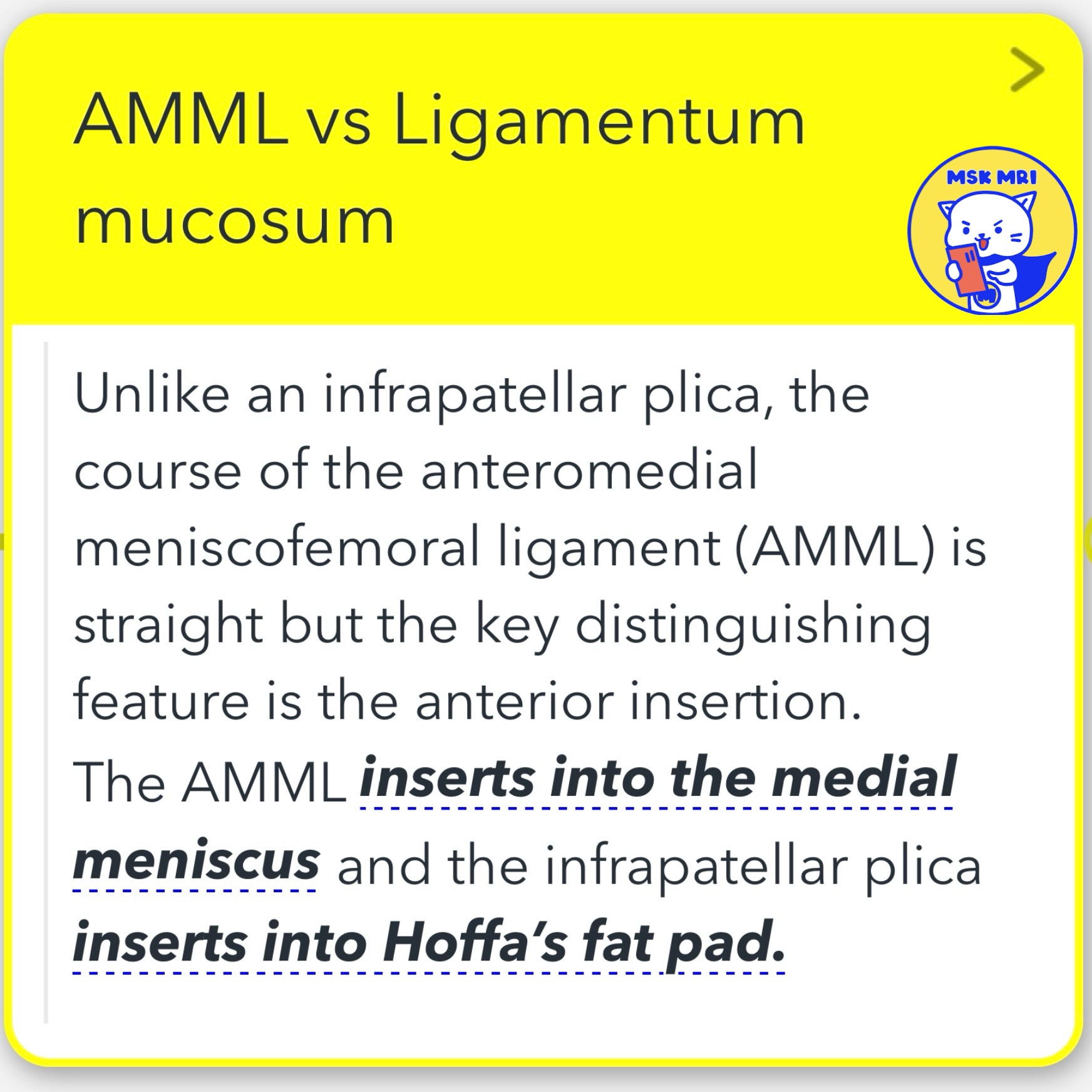
Comparison of Anteromedial Meniscofemoral Ligament and Ligamentum Mucosum
1️⃣ Anteromedial Meniscofemoral Ligament (AMML):
- Definition: An uncommon accessory ligament that extends from the anterior horn of the medial meniscus, running parallel to the Anterior Cruciate Ligament (ACL), to insert into the medial wall of the lateral femoral condyle adjacent to the ACL insertion.
- Location and Course: The AMML is noted for its straight course, distinguishing it from other ligamentous structures within the knee, with a specific anterior insertion into the medial meniscus.
2️⃣ Ligamentum Mucosum (Infrapatellar Plica):
- Definition: A centrally located synovial fold within the knee, extending from the inferior pole of the patella through Hoffa’s fat pad and to the intercondylar notch, paralleling the ACL.
- Plicae Overview: The knee contains three main plicae - suprapatellar, infrapatellar (Ligamentum Mucosum), and medial patellar, which are synovial folds that play roles in knee structure and function.
📌 Key Differences:
- Course and Insertion: The primary difference between AMML and Ligamentum Mucosum lies in their course and points of insertion. AMML is distinguished by its straight course and anterior insertion into the medial meniscus, whereas the Ligamentum Mucosum (infrapatellar plica) inserts into Hoffa’s fat pad, with a central location that runs through the knee.
"Visualizing MSK Radiology: A Practical Guide to Radiology Mastery"
© 2022 MSK MRI Jee Eun Lee All Rights Reserved.
#VisualizingMSK #aclanatomy #ACL #plica #accessoryligament
You may not distribute or commercially exploit the content.
Nor may you transmit it or store it on any other website or other forms of the electronic retrieval system.


