Click the link to purchase on Amazon 🎉📚
==============================================
🎥 Check Out All Videos at Once! 📺
👉 Visit Visualizing MSK Blog to explore a wide range of videos! 🩻
https://visualizingmsk.blogspot.com/?view=magazine
📚 You can also find them on MSK MRI Blog and Naver Blog! 📖
https://www.instagram.com/msk_mri/
Click now to stay updated with the latest content! 🔍✨
==============================================
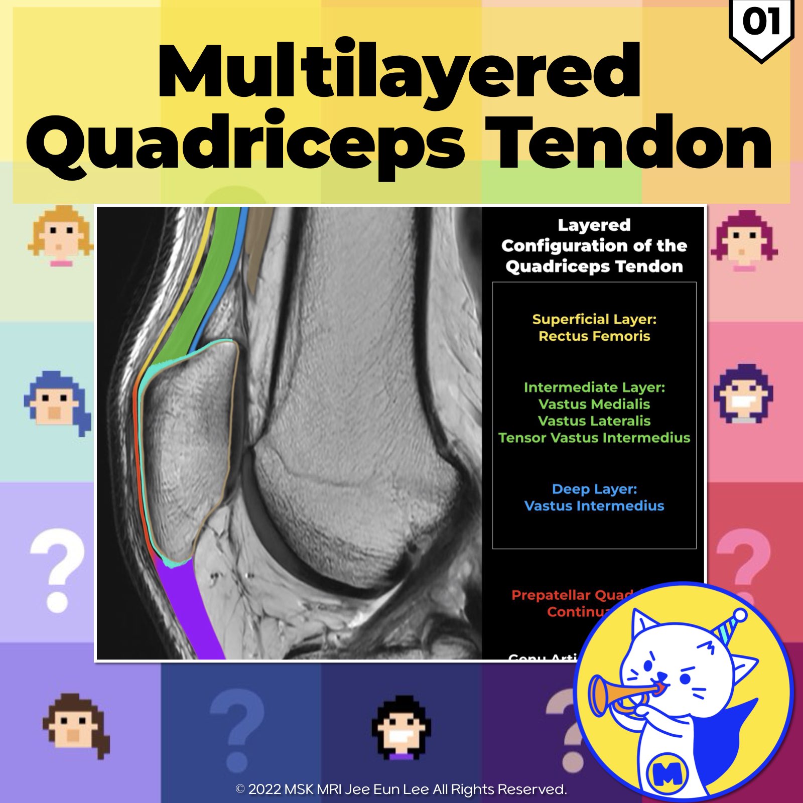
📌 Overview of Quadriceps Tendon:
The quadriceps tendon has a unique laminated appearance on sagittal MR images, with low-signal intensity bands separated by linear fat.
It can appear in different configurations: trilaminar (56%), bilaminar (30%), or quadrilaminar (8% )
1️⃣ Most Superficial Layer:
- Originates from the rectus femoris muscle.
2️⃣ Intermediate Layer:
- Formed by the fusion of the tendons of the vastus lateralis and vastus medialis.
- Attaches to the anterosuperior surface of the proximal patella, posterior to the rectus femoris insertion.
- Vastus lateralis tendon is anterior to vastus medialis tendon in the thigh, converging at the patella【RadioGraphics 2018; 38:2069–2101】.
3️⃣ Deepest Layer:
- Comprised of the vastus intermedius tendon, which inserts into the anterosuperior surface of the patella.
✅ Additional Structures:
1️⃣ Tensor of Vastus Intermedius (TVI):
- Newly discovered muscle in the intermediate layer.
- Located between the vastus lateralis and vastus intermedius.
- Merges with a broad, flat aponeurosis before attaching to the medial patella.
2️⃣ Prepatellar Quadriceps Continuation:
- Consists of rectus femoris tendon fibers connecting the quadriceps and patellar tendons.
- Very thin and adherent to the patella, making differentiation from the patellar cortex challenging.
3️⃣ Articularis Genus (Articular Muscle of the Knee):
- Originates from the distal femur.
- Seen on sagittal MR imaging in most patients.
- Stabilizes the suprapatellar recess and joint capsule during extension.
- Deepest component of the extensor mechanism, inserting on the suprapatellar pouch
References:
- RadioGraphics 2018; 38:2069–2101
- Magn Reson Imaging Clin N Am 22 (2014) 601–620
- Knee Surg Sports Traumatol Arthrosc. 2018 Mar;26(3):727-738
- Knee Surg Sports Traumatol Arthrosc. 2018 Mar;26(3):727-738
- AJR 2009; 192:W111–W116
"Visualizing MSK Radiology: A Practical Guide to Radiology Mastery"
© 2022 MSK MRI Jee Eun Lee All Rights Reserved.
No unauthorized reproduction, redistribution, or use for AI training.
#QuadricepsTendon, #SagittalMRI, #MusculoskeletalImaging, #RectusFemoris, #VastusLateralis, #VastusMedialis, #VastusIntermedius, #TensorVastusIntermedius, #PrepatellarQuadriceps, #ArticularisGenus


'✅ Knee MRI Mastery > Chap 4BCD. Anterior knee' 카테고리의 다른 글
| (Fig 4-B.06) Symptomatic Bipartite Patella (1) | 2024.06.10 |
|---|---|
| (Fig 4-B.05) Patellar Comparison/ Multipartite, Bipartite, Vertical Fracture (0) | 2024.06.10 |
| (Fig 4-B.04) Bipartite Patella and Saupe Classification (1) | 2024.06.10 |
| (Fig 4-B.03) Retropatellar Articular Surface and Wiberg Classification (0) | 2024.06.10 |
| (Fig 4-B.02) Anatomy of the Multilayered Quadriceps Tendon/ Part 2 (0) | 2024.06.09 |