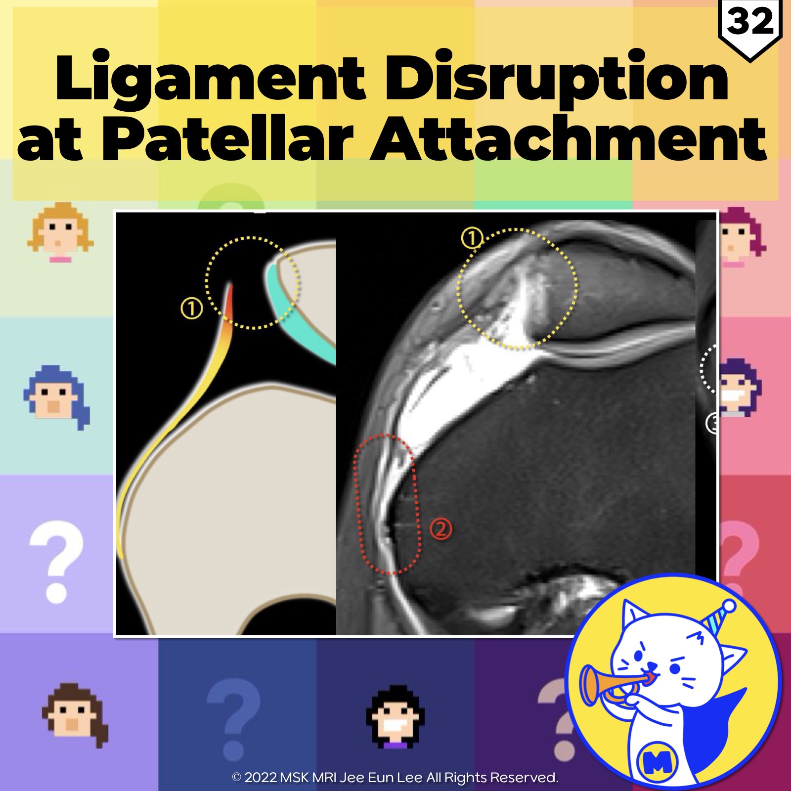
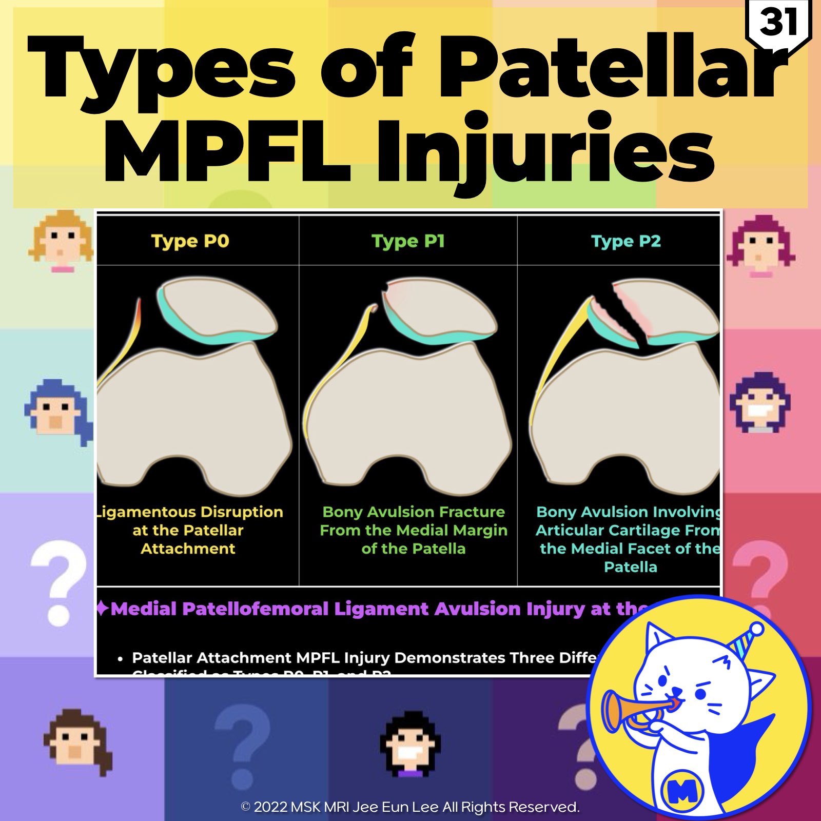
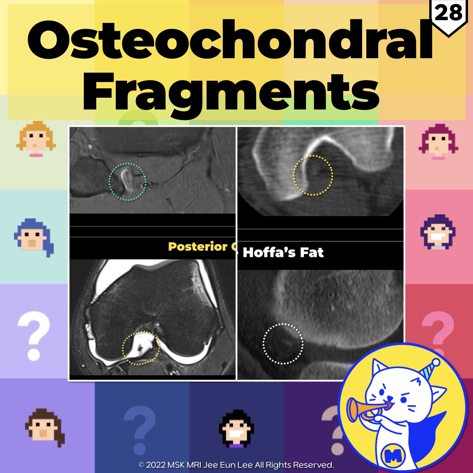
본문 바로가기

분류 전체보기608

(Fig 1-A.06) Medial Meniscus Posterior Root, Anatomy, degeneration, tear https://youtu.be/GvR9ULGrDOw https://youtu.be/kEuMzUrHD0M Category Description Insertion Points - Inserts along the downward slope of the intercondylar fossa, anterior to the posterior cruciate ligament. - Extends from the tibial attachment point to just lateral from the articular cartilage inflection point of the medial tibial plateau. - Commences just lateral to the apex of the medial tibial p.. 2024. 1. 5.
(Fig 1-A.04) Anatomy of lateral Meniscus anterior root, tibial spine fracture https://youtu.be/w2QemJqJBfsTitle: Unraveling the Complexities of the Lateral Meniscus and ACL on MRI https://youtu.be/kEuMzUrHD0M 🔍 Deep Dive into Anatomy and Injury: the intricate relationship between the anterior root of the lateral meniscus (LM) and the anterior cruciate ligament (ACL) in the knee, as revealed by MRI. Unique Structure: The anterior root of the LM is distinct with its fan-sha.. 2024. 1. 5.
Anatomy of Medial Meniscus anterior root, variation https://youtu.be/dECIy2gvacM https://youtu.be/kEuMzUrHD0M The medial meniscus has an anterior root that attaches to the area in front of the tibial attachment of the ACL, specifically in the intercondylar fossa of the tibia. Additionally, the posterior fibers of the anterior horn of the medial meniscus connect to the transverse ligament. This anterior root can vary in attachment, with most peopl.. 2024. 1. 4.
(Fig 1-A.02) Comparison of Medial and Lateral Meniscus https://youtu.be/Q1SWxmc7sBA https://youtu.be/kEuMzUrHD0M 2024. 1. 2.
MPFL (Medial Patellofemoral Ligament) Anatomy Medial Patellar Retinacular Structures✓ Medial patellofemoral ligament (MPFL): (most important component) ✓ Medial patellotibial ligament (MPTL) ✓ Medial patellomeniscal ligament (MPML) ✓ Medial quadriceps tendon femoral ligament (MQTFL) Composition of the MPFL A stronger transverse band and a more variable, weaker oblique decussation. Two components: Transverse Component (tMPFL) Originates more.. 2023. 12. 31.
Dislocation of Polyethylene Insert Dislocation of a polyethylene insert Mechanism of Dislocation (DPI in TKA) Proposed Cause Atraumatic nature No clear cause identified Traumatic mechanism (insert dislocation) Repeated posteriorly directed compressive force on the polyethylene insert from the femur Biomechanical basis for dislocation During knee flexion, femoral tibial contact moves posteriorly, leading to anterior lift-off of th.. 2023. 12. 30.
Polyethylene wear–induced synovitis Polyethylene Wear Polyethylene components used in total knee arthroplasty (TKA) can experience wear on the articular surfaces due to abrasion and fatigue This wear generates polyethylene debris, which triggers an inflammatory response. Over time, this response can lead to osteolysis and loosening. Radiographic Findings of Polyethylene Wear Progressive joint space narrowing Sequential radiographs.. 2023. 12. 29.
[Case_Adv] Accessory Iliotibial Band-Meniscal Ligament, normal variant, lateral meniscus https://youtu.be/wmpIaimU2gE The anatomic variation of the anterolateral fixation of the lateral meniscus to the ITB, which we have termed the “accessory iliotibial band–meniscal ligament” or “AIML.” An AIML was present in 13.3% of all patients (136/1019). AJR Am J Roentgenol. 2019 Oct;213(4):912-917. Although the attachments of the anterior and posterior horn are relatively consistent, the ante.. 2023. 2. 2.
[Elbow-01] Acute gouty arthritis https://youtube.com/shorts/EXt4aC9NvpUzA Acute gout is intensely inflammatory, presenting as a relatively sudden onset severe pain in a joint associated with swelling, warmth, and redness that may extend beyond the joint, mimicking cellulitis. The earliest episode of acute gout are usually mono-articular and usually affect, in decreasing order of frequency, the first metatarsophalangeal joint (‘.. 2022. 12. 27.
[Knee-04] Anatomy of layers and components, Posterolateral corner 01 https://youtu.be/lMx9cjkzNZw Three structural layers Layer 1:most superficial Fascia lata lliotibial tract with its anterior expansion Superficial portion of biceps femoris with its posterior expansion ​ Layer 2 Quadriceps retinaculum anteriorly Two patellofemoral ligaments or retinacula posteriorly Lavers 1 and 2 merges at lateral aspect of the patella ​ Layer 3:deepest layer The lateral joint .. 2022. 12. 25.
[Knee-03] Tear of Popliteus tendon, Posterolateral corner (PLC) injury https://youtu.be/XVRwYJa4f2M 1. Tear of the popliteus tendon A. Posterolateral (arcuate) complex LCL Popliteus tendon Lateral head of gastrocnemius muscle Arcuate ligament Popliteofibular ligament Fabellofibular ligament Stoller's Orthopaedics and Sports Medicine: The Knee B. Posterolateral corner (PLC) injury Injury to a component of the lateral collateral ligament complex in association with a.. 2022. 12. 24.
[Knee-02] Popliteus tendon anatomy, Posterolateral attachment https://youtu.be/pkdL_HwgPi0 Popliteus The popliteus musculotendinous complex—consisting of the popliteus tendon, popliteus muscle, and PFL The tendon, which is intracapsular, but extra-articular and extrasynovial, ascends around the posterolateral aspect of the knee The tendinous attachment lies anteroinferior to the proximal attachment of the lateral collateral ligament on the lateral epicondy.. 2022. 12. 24.
[Knee-01] Tear of anteroinferior popliteomeniscal fascicle, posterolateral corner injury https://youtu.be/he2-XqAyvaA * The popliteomeniscal fascicles arise from the periphery of the posterior horn of the lateral meniscus and form a posterolateral meniscocapsular extension, which creates the popliteus hiatus. * The fascicles not only form the hiatus, which allows the popliteus tendon an avenue to course intra-articularly and maintain the integrity of the joint but stabilize and cont.. 2022. 12. 21.
[Wrist_02] Scapholunate ligament tear and Dorsal capsulo-scapholunate septum tear https://youtu.be/R_DCrdBY9Pg https://www.instagram.com/msk_mri/ MSKMRI(@msk_mri) • Instagram 사진 및 동영상 팔로워 1,850명, 팔로잉 84명, 게시물 194개 - MSKMRI(@msk_mri)님의 Instagram 사진 및 동영상 보기 www.instagram.com The scapholunate ligament (SLL) is a C-shaped structure, and has 3 histologically and functionally distinct components, dorsal, volar and proximal (interosseous) components. The dorsal part of the scapholu.. 2022. 12. 20.
[Ankle and Foot_04] Dorsal calcaneocuboid joint injury, Peroneus brevis tendon tear https://www.instagram.com/msk_mri/ The calcaneocuboid joint is stabilized by the dorsal calcaneocuboid ligament, short and long plantar ligaments, and calcaneocuboid component of the bifurcate ligament. ​​The calcaneocuboid joint capsule is reinforced dorsolaterally by the dorsal calcaneocuboid ligament The long and short plantar ligaments (sometimes referred to together as the plantar or inferi.. 2022. 12. 18.
[Wrist_01] Madelung deformity, carpal instability adaptive, Vicker’s ligament The commonly used Mayo classification divides carpal instability into four major categories. 1. Carpal instability dissociative (CID), where there is dissociation of the bones in a single carpal row. The proximal carpal row is most commonly affected (i.e., SL instability; lunotriquetral instability). Distal carpal row dissociation is rare. 2. Carpal instability nondissociative includes instabili.. 2022. 12. 18.
[Shoulder_02] Rotator cuff footprint, Articular-delaminated partial-thickness tear of the infraspinatus tendon Articular-surface partial-thickness tear with intratendinous horizontal splitting tear of the infraspinatus tendon Intact supraspinatus tendon The footprint of the supraspinatus on the greater tuberosity is much smaller than previously believed, and this area of the greater tuberosity is actually occupied by a substantial amount of the infraspinatus. The anterior part of the infraspinatus partia.. 2022. 12. 18.
[Shoulder_01] Rotator cuff footprint, supraspinatus, and infraspinatus tendons Relatively small, triangular, the anteromedial footprint of the supraspinatus tendon at the superior facet and a comparatively large, trapezoidal footprint of the infraspinatus tendon occupying the anterolateral portion of the superior facet and the entire middle facet. The footprint of the supraspinatus had a triangular shape, which tapered away from the joint capsule. The footprint of the infr.. 2022. 12. 18.
[Ankle and Foot_02] Two fascicles of the anterior talofibular ligament (ATFL) The anterior talofibular ligament (ATFL) is composed of the superior (S-ATFL) and inferior fascicle (I-ATFL). Both fascicles have a contiguous footprint on the anterior distal fibula border, however, their distal insertion on the talus is located apart from each other. The superior fascicle of the ATFL is a distinct anatomical structure, whereas the inferior ATFL fascicle and the CFL share some .. 2022. 12. 12.
[Ankle and Foot_01] anterior talofibular ligament (ATFL), and posterior talofibular ligament (PTFL) The lateral ligament complex of the ankle consists of the anterior talofibular ligament (ATFL), calcaneofibular ligament (CFL), and posterior talofibular ligament. This ligament plays an important role in limiting anterior displacement, internal rotation of the talus, and plantar flexion of the ankle The anterior talofibular ligament extends from the anterolateral malleolar tip to the talar neck.. 2022. 12. 11.
[PCL 07] 02. Popliteus musculotendinous complex, popliteomeniscal fascicle, popliteal bursa, cyamella With popliteus tears, it is important to note - the site of the tear (intra- articular or extra-articular), - the extent of tear (partial or full-thickness), - the presence of a bone fragment if there has been tendon avulsion - degree of tendon retraction Partial-thickness tear of the popliteus tendon, High-grade partial tearing of the PT at its femoral insertion Most popliteus tears are extra-a.. 2022. 11. 19.